
News & Scientific Highlights

World record in time-resolved tomography
Researchers from the Helmholtz Zentrum Berlin (HZB) and the TOMCAT beamline have achieved a new world record in time-resolved tomography by measuring over 200 tomographies per second during heating of an evolving aluminium metal foam.

High-numerical-aperture optics is key to ultra-fast tomographic microscopy
A novel high-numerical-aperture macroscope optics dedicated to high-temporal and high-spatial resolution X-ray tomographic microscopy is available at TOMCAT. Coupled with the in-house developed GigaFRoST camera, this highly efficient imaging setup enables tomographic microscopy studies at 20 Hz and beyond, opening up new possibilities in tomographic investigations of dynamic processes. A detailed characterization of the macroscope performance was published in Journal of Synchrotron Radiation on May 21, 2019.

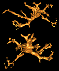
From whole organ imaging down to single cell analysis
Researchers from the TOMCAT beamline, University College London (UCL), IDIBAPS and Universitat Pompeu Fabra (UPF) have developed a methodology that allows the multiscale analysis of the structural changes resulting from remodelling cardiovascular diseases, from whole organ down to single-cell level. This methodology has been published as an article in the journal Scientific Reports on May 6th 2019.

PSI Thesis Medal goes to Dr. Matias Kagias
The PSI Thesis Medal is awarded every second year to the best PhD thesis performed at the Paul Scherrer Institut. Matias received the prize for his excellent thesis entitled "Direct Self-Imaging Methods for X-ray Differential Phase and Scattering Imaging". Congratulations!

Virtual lens improves X-ray microscopy
A method developed by PSI researchers makes X-ray images of materials even better. The researchers took a number of individual images while moving an optical lens. From these, with the help of computer algorithms, they generated one overall image.
Soft-tissue evidence for homeothermy and crypsis in a Jurassic ichthyosaur
Synchrotron-based X-ray tomographic microscopy of melanophores (skin pigment cells) of an amazingly well preserved 180 million years old ichtyosaur (extinct marine reptile similar to whales) contributed in a multidisciplinary investigation to the new findings published today in Nature.

TOMCAT paper on hard X-ray multi-projection imaging published
The TOMCAT team in collaboration with scientists from CFEL, MaxIV and ESRF developed a method for hard X-ray multi-projection imaging, using a single crystal to split the beam into multiple beams with different directions.

PSI spin-off GratXray wins Swiss Technology Award 2017
A spin-off from PSI has received this year's Swiss Technology Award: The young company GratXray is developing a new method for early diagnosis of breast cancer.
New TOMCAT paper: The GigaFRoST camera and readout system
The PSI in-house developed GigaFRoST high-speed camera and readout system is available for fast imaging experiments at the TOMCAT beamline, opening up exciting new possibilities for the observation of fast dynamic phenomena with X-ray tomography.