
Scientific Highlights
X-band prototype structure
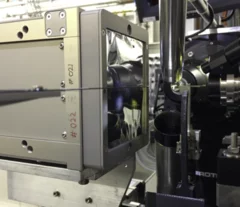
Radio-frequency structures at X-band frequencies (~ 12 GHZ) are being considered for applications in compact Free Electron Lasers, medical linacs, a future linear collider (CLIC project) and as a diagnostic for measuring ultra-short (femtosecond) electron pulses in FELs. A first prototype of such a structure has been built at PSI employing the realization procedures that have been developed for the C-Band (6 GHz) structures of the SwissFEL linac.
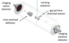
Breaking Through the False Coincidence Barrier in Electron–Ion Coincidence Experiments
The false coincidence background has so far limited the analytical application of PEPICO, photoelectron photoion coincidence. A new photoioin rastering technique has been developed to separate the wheat from the chaff and identify true coincidences based on the ion hit time and position. This expands the dynamic range of the experiment by at least two orders of magnitude, allowing for novel applications to look for reactive intermediates and short lived species in reaction environments.
Structure and Conductivity of Epitaxial Thin Films of In-Doped BaZrO3‑Based Proton Conductors
Epitaxial thin films of the proton-conducting perovskite BaZr0.53In0.47O3−δH0.47−2δ, grown by pulsed laser deposition, were investigated in their hydrated and dehydrated conditions through a multitechnique approach with the aim to study the structure and proton concentration depth profile and their relationship to proton conductivity.
EUCALL finishes first year, bearing new technologies
The European Cluster of Advanced Laser Light sources (EUCALL), a European Union-funded project that aims to foster links between accelerator- and laser-driven X-ray facilities, has completed the first year of its three year project period. The project successfully met all twenty of its milestones for the year, producing a new open-source tool for experiment simulations and developing specifications for several pieces of new scientific equipment.

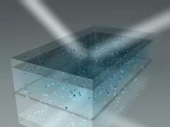
100 Hz neutron radiography at the BOA beamline using a parabolic focussing guide
The recent developments in scientific complementary metal oxide semiconductor (sCMOS) detector technology allow for imaging of relevant processes with very high temporal resolution with practically negligible readout time. However, it is neutron intensity that limits the high temporal resolution neutron imaging. In order to partially overcome the neutron intensity problem for the high temporal resolution imaging, a parabolic neutron focussing guide was utilized in the test arrangement and placed upstream the detector in such a manner that the focal point of the guide was positioned slightly behind the scintillator screen. In such a test arrangement, the neutron flux can be increased locally by about one order of magnitude, albeit with the reduced spatial resolution due to the increased divergence of the neutron beam. In a pilot test application, an in-situ titration system allowing for a remote delivery of well-defined volumes of liquids onto the sample stage was utilized. The process of droplets of water (H2O) falling into the container filled with heavy water (D2O) and the subsequent process of the interaction and mixing of the two liquids were imaged with temporal resolution of 0.01 s.

Progress in High-resolution Neutron Imaging at the Paul Scherrer Institut – The Neutron Microscope Project
The recent improvement on the capability of neutron imaging that allows acquiring neutron images with isotropic spatial resolution of about 5 micrometres is demonstrated. This is achieve by combining the tailor-made high-numerical aperture magnifying optics together with a thin isotopically-enriched 157Gd2O2S:Tb scintillator screens (see Trtik & Lehmann, NIM-A 788 (2015) 67-70). The newly achieved level of the spatial resolution represents about 30% enhancement compared to the first prototype (see Trtik et al, Physics Procedia 69 (2015) 169-176) and approximately six-fold enhancement in the spatial resolution capabilities available for the general users community at PSI before the start of the Neutron Microscope project.
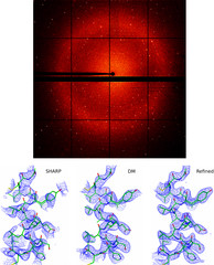
First protein structure solved using the JUNGFRAU detector
JUNGFRAU is a charge-integrating, two-dimensional pixel detector developed at the Paul Scherrer Institut for use at free-electron lasers, in particular SwissFEL, and synchrotron light sources. On the 10th October, the first protein crystallography experiment using the JUNGFRAU detector, was performed at the beamline X06SA (PXI) of the Swiss Light Source by the members of the Protein Crystallography and Detectors groups at PSI.
C–H Activation on Co,O Sites: Isolated Surface Sites versus Molecular Analogs
The activation and conversion of hydrocarbons is one of the most important challenges in chemistry. This work shows that isolated Co(II) sites are catalysts for a number of hydrocarbon conversion reactions, such as the dehydrogenation of propane, the hydrogenation of propene, and the trimerization of terminal alkynes. The data are consistent with all of these reactions occurring by a common mechanism, involving heterolytic C–H or H–H activation via a 1,2 addition across a Co–O bond.
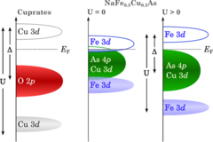
Novel insulating phase in iron-pnictide materials
The first example of an insulating phase which is close to the superconducting phase in an iron-pnictide system has been recently observed in heavy Cu-doped NaFe1-xCuxAs (x > 0.3). A combined study by angle-resolved photoemission spectroscopy (ARPES) and density functional theory (DFT) calculations revealed that on-site Coulomb repulsion and enhanced Hund’s rule coupling are responsible for the insulating behavior. The results show that the insulating phase in NaFe0.5Cu0.5As resembles the situation in the parent compounds of the high-Tc cuprate superconductors.
First protein structure solved using the JUNGFRAU detector!
JUNGFRAU is a charge-integrating, two-dimensional pixel detector developed at the Paul Scherrer Institut for use at free-electron lasers, in particular SwissFEL, and synchrotron light sources. On the 10th October, the first protein crystallography experiment using the JUNGFRAU detector, was performed at the beamline X06SA (PXI) of the Swiss Light Source by the members of the Protein Crystallography and Detectors groups at PSI.