
News & Scientific Highlights
Fast scanning coherent X-ray imaging using Eiger
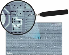
The smaller pixel size, high frame rate, and high dynamic range of next-generation photon counting pixel detectors expedites measurements based on coherent diffractive imaging (CDI). The latter comprises methods that exploit the coherence of X-ray synchrotron sources to replace imaging optics by reconstruction algorithms. Researchers from the Paul Scherrer Institut have recently demonstrated fast CDI image acquisition above 25,000 resolution elements per second using an in-house developed Eiger detector. This rate is state of the art for diffractive imaging and even on a par with the fastest scanning X-ray transmission instruments. High image throughput is of crucial importance for both materials and biological sciences for studies with representative population sampling.

X-ray tomography reaches 16 nm isotropic 3D resolution
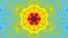
Researchers at PSI reported a demonstration of X-ray tomography with an unmatched isotropic 3D resolution of 16 nm in Scientific Reports. The measurement was performed at the cSAXS beamline at the Swiss Light Source using a prototype instrument of the OMNY (tOMography Nano crYo) project. Whereas this prototype measures at room temperature and atmospheric pressure, the OMNY system, to be commissioned later this year, will provide a cryogenic sample environment in ultra-high vacuum without compromising imaging capabilities. The researchers believe that such a combination of advanced imaging with state-of-the-art instrumentation is a promising path to fill the resolution gap between electron microscopy and X-ray imaging, also in case of radiation-sensitive materials such as polymer structures and biological systems.

Unique insight into carbon fibers on the nanoscale
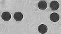
The investigation of the mechanical properties of carbon fibers benefits from highly resolved three-dimensional density maps within representative volumes, but such images are not easily obtained with standard methods. Scientists from the Paul Scherrer Institut in collaboration with Honda R&D in Germany have recently visualized density distributions on the sub-micrometer scale within entire carbon fiber sections, revealing surprising graphite distributions within the fibers. This capability will prove useful for the systematic characterization of fibers, contributing to the development of light and robust materials at lower costs.
Röntgen-Laser: Auf dem Weg zur Strukturbestimmung von Nanoteilchen
An Freie-Elektronen-Röntgen-Lasern wie dem zukünftigen SwissFEL des Paul Scherrer Instituts (PSI) sollen unter anderem die Strukturen von komplexen Nanoteilchen bis hin zu Biomolekülen untersucht werden. Dabei ist nicht nur die eigentliche Messung eine Herausforderung, sondern auch die Rekonstruktion der Struktur aus den Messdaten. Forscher des PSI haben nun einen optimierten mathematischen Weg aufgezeigt, wie man aus so gewonnen Messdaten eine deutlich bessere Auflösung bei der Bestimmung der Struktur eines einzelnen Teilchens erhält. Das Verfahren wurde an der Synchrotron Lichtquelle Schweiz des PSI erfolgreich getestet.Cette actualité n'existe qu'en allemand.
Imager des échantillons qui fluctuent à l'aide de rayons X
Les rayons X sont utilisés pour inspecter la structure à l'échelle nanométrique d'objets variés comme des cellules biologiques ou des dispositifs de stockage magnétiques. L'imagerie à très haute résolution impose cependant de très fortes contraintes, autant sur l'appareil que sur l'échantillon lui-même. Des chercheurs à la Technische Universität München et au PSI viennent de démontrer comment ces conditions peuvent être relaxées sans perte de qualité d'image. Ils ont de plus montré comment la même approche permet d'imager des échantillons qui fluctuent très rapidement, comme les matériaux magnétiques utilisés pour le stockage de données.
Nanoforscher untersuchen Karies
Forscher der Universität Basel und des Paul Scherrer Instituts konnten im Nanomassstab zeigen, wie sich Karies auf die menschlichen Zähne auswirkt. Ihre Studie eröffnet neue Perspektiven für die Behandlung von Zahnschäden, bei denen heute nur der Griff zum Bohrer bleibt. Die Forschungsergebnisse wurden in der Fachzeitschrift «Nanomedicine» veröffentlicht.Cette actualité n'existe qu'en allemand.
Röntgen-Methode hilft Hirnerkrankungen besser zu verstehen
Ein internationales Forschungsteam hat eine neue Methode entwickelt, mit der man detaillierte Röntgenbilder von Hirngewebe erstellen kann. Die Methode wurde verwendet, um die Myelinscheide der Nervenfasern sichtbar zu machen. Schäden an der Myelinscheide führen zu verschiedenen Erkrankungen wie etwa Multiple Sklerose. Die Anlage, an der diese Aufnahmen erstellt werden können, wird an der Synchrotron Lichtquelle Schweiz SLS des Schweizer Paul Scherrer Instituts betrieben.Cette actualité n'existe qu'en anglais et allemand.
Fortschritt für die Knochen-Forschung
Hochauflösendes Verfahren zur Nano-Computertomographie entwickeltEin neuartiges Nano-Tomographie-Verfahren, das von einem Team der TU München, des Paul Scherrer Instituts (PSI) und der ETH Zürich entwickelt wurde, erlaubt erstmals computertomographische Untersuchungen feinster Strukturen mit einer Auflösung im Nanometerbereich. Mit Hilfe der neuen Methode können etwa dreidimensionale Innenansichten fragiler Knochenstrukturen erstellt werden.Cette actualité n'existe qu'en anglais et allemand.