The broad application of lithium ion batteries (LIB), from portable electronics to electric vehicles, motivates the urgent search for higher performance and improved safety and lifetime. Lithium-rich nickel, manganese and cobalt oxide (Li-rich NMC) offers performance advantages over industry standards, including an energy density of over 900 Wh/kg, higher than that of LiFePO4 or LiCoO2 materials. However, processes such as first-cycle activation and repeated lithiation/delithiation result in chemical and morphological degradation of the material, impeding its commercialization. Extensive studies were carried out to understand the aging mechanism of Li-rich NMC in recent years, mainly on the primary particle level (50-100 nm grains) or on the bulk level. In this work we study the morphological and crystalline structural changes of secondary particles (5-20 um spheres), offering insight into the battery material from a different perspective on a different scale. The research was carried out as a collaboration between cSAXS beamline (X12SA), the microXAS beamline (X05LA), and the Electrochemistry Laboratory, Paul Scherrer Institute, Switzerland.
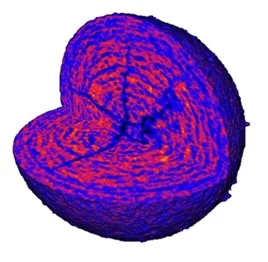
The 3D information on the material morphology was retrieved via ptychographic X-ray computed tomography (PXCT), a high-resolution lensless coherent diffractive imaging method, that offers an estimated 3D resolution down to 35 nm for these particles. Dozens of secondary particles were imaged, including those from the pristine stage, after first delithiation, and after a few months of cycling. Micro-cracks were observed on multiple particles, as shown in the figure, suggesting the stage, the spatial extent, and the particle-size dependency of the morphological degradation. Subsequent to PXCT experiments, a series of scanning X-ray diffraction microscopy (SXDM) measurements were performed on the same samples to examine the crystallographic structure and its correlation to the observed morphology. Interestingly, the SXDM results present an expected spatially-dependent crystal structure, seen as core-shell, inviting for further studies. With ongoing instrumentation advances, future studies will also seek to image cells operando. The project was supported by the Swiss National Science Foundation (SNSF) grant number 200021_152554 and 200020_169623.
Contact
Dr. Manuel Guizar-Sicairos
Beamline Scientist, 6113, WLGA/227
Paul Scherrer Institut
Telephone: +41 56 310 3409
E-mail: manuel.guizar-sicairos@psi.ch
Original Publication
Correlated X-Ray 3D Ptychography and Diffraction Microscopy Visualize Links between Morphology and Crystal Structure of Lithium-Rich Cathode Materials
Esther H.R. Tsai, Juliette Billaud, Dario F. Sanchez, Johannes Ihli, Michal Odstrčil, Mirko Holler, Daniel Grolimund, Claire Villevieille, and Manuel Guizar-Sicairos
iScience 11, 356-365 (2019) Date
DOI: 10.1016/j.isci.2018.12.028