Aller au contenu principal
  • Paul Scherrer Institut PSI
  • PSI Research, Labs & User Services

Digital User Office

  • Digital User Office
  • DE
  • EN
  • FR
Paul Scherrer Institut (PSI)
Rechercher
Paul Scherrer Institut (PSI)

Hauptnavigation

  • Research at PSIOuvrir ce point de menu principal
    • Research Initiatives
    • Ethics and Research integrity
    • Scientific Highlights
    • Scientific Events
    • Scientific Career
    • PSI-FELLOW
    • PSI Data Policy
  • Research Divisions and LabsOuvrir ce point de menu principal
    • Overview
    • Research with Neutrons and Muons
    • Photon Science
    • Energy and Environment
    • Nuclear Energy and Safety
    • Biology and Chemistry
    • Scientific Computing, Theory and Data
    • Large Research Facilities
  • Facilities and InstrumentsOuvrir ce point de menu principal
    • Overview
    • Large Research Facilities
    • Facilities
    • PSI Facility Newsletter
  • PSI User ServicesOuvrir ce point de menu principal
    • User Office
    • Methods at the PSI User Facilities
    • Proposals for beam time
    • Proposal Deadlines
    • Data Analysis Service (PSD)
    • EU support programmes
  • New ProjectsOuvrir ce point de menu principal
    • SLS 2.0
    • IMPACT
  • DE
  • EN
  • FR

Digital User Office (mobile)

  • Digital User Office

Vous êtes ici:

  1. PSI Home
  2. Labs & User Services
  3. PSD
  4. LSB
  5. CXS
  6. Research

Navigation secondaire

Coherent X-Ray Scattering Group

  • People
  • Research
  • Scientific Highlights and News
  • Publications

Info message

Ce contenu n'est pas disponible en français.

Research


smeulders.jpg

Small-angle x-ray scattering (SAXS)

The small angle diffraction of hard x-rays provides statistical information on nanoscale ordering and structure. We develop experimental methods for time resolved measurement of structural changes induced by laser radiation, microfluidic mixing or temperature changes. Image on the left shows interlocked octameric rings of CS2 hydrolase (abstract in journal webpage).

2015 Liebi bone.jpg

Scanning SAXS in 2D and 3D

By scanning the sample relative to the x-ray beam we obtain spatially resolved SAXS datasets. For each point in the images a statistical description of ordering in the nanoscale is available. This technique can be further combined with sample rotations to probe ordering in 3D or to obtain measurements that are resolved in three-dimensions via tensor computed tomography (CT). In the image on left we show the result on a trabecular bone fragment of roughly two and a half millimetre long. The image shows a spatially resolved representation of the arrangement, orientation, and degree of orientation of nanoscale collagen fibrils (abstract in journal webpage).

ptychotomo.jpg

Ptychography - Scanning x-ray diffraction microscopy

Using iterative algorithms as computational lenses coherent x-ray diffraction patterns are phased and render quantitative phase and amplitude images of the sample under study. We investigate and develop sample environments, measurement schemes, and reconstruction algorithms to further the development and application of this technique. Shown on the left is a 3D quantitative reconstruction of a small section of mouse femur bone, highlighting the osteocyte lacunae (L) and the connecting canaliculi network (C) (abstract in journal webpage).

igp 240 igp 578x527  Ta2O5.png

OMNY - tOMography Nano crYo stage

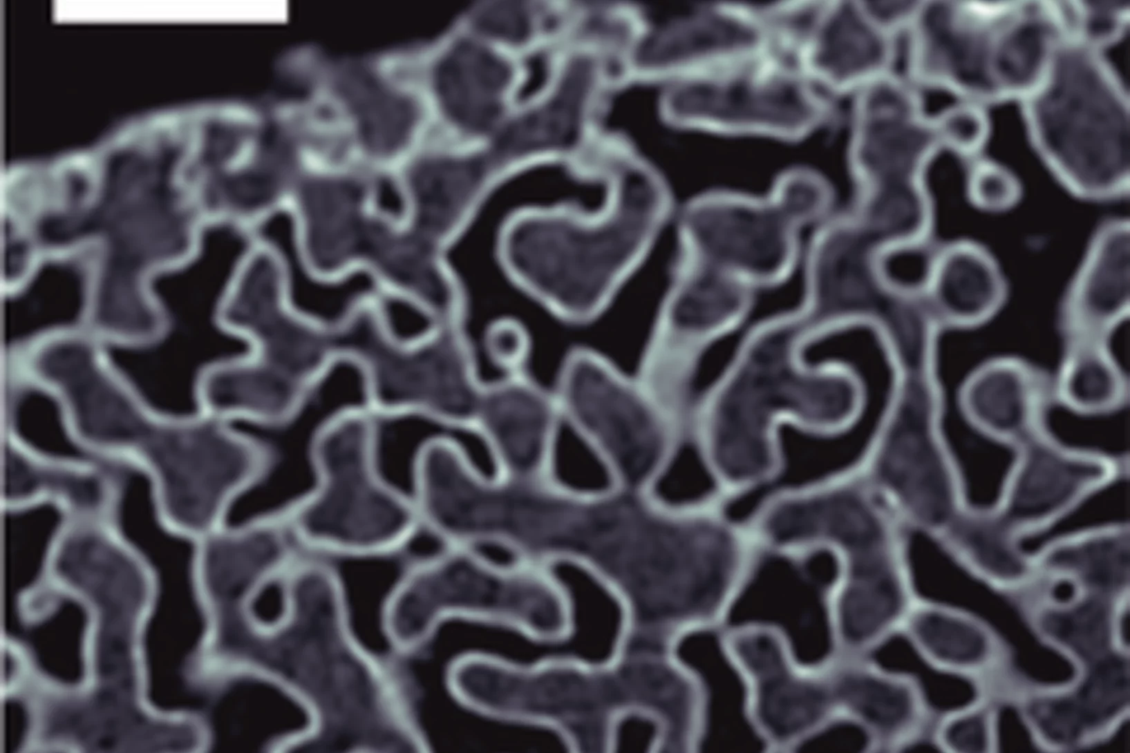
We work closely to the OMNY project in developing and testing a dedicated instrument for high-resolution scanning tomography based on differential interferometry. Two instruments exist, one in air and room temperature for added flexibility in the measurement environment, and a second one in vacuum and cryogenic temperatures to allow measurements on radiation sensitive materials such as soft tissue. This method achieves a stability of better than 10 nm between beam defining optics and sample. As an example we show on the left a tomogram of a coated nanoporous glass sample where three distinct gray levels are visible for air (black), glass (gray), and Ta2O5 (white). The projections were measured using ptychography and an isotropic 3D resolution of 16 nm was achieved (abstract in journal webpage).

FZP wavefront.jpg

Characterization of focused beams and X-ray wavefronts

Coherent X-ray beams can be accurately characterized using ptychography. Upon scanning a test sample across the beam and measuring the resulting far-field diffraction patterns, reconstruction algorithms for ptychography retrieve phase and amplitude of the sample and the incoming beam. The reconstructed illumination can be numerically propagated to virtually any plane along the propagation axis. On the left we show an example of such characterization where a 23 nm beam focused by a Fresnel zone-plate (FZP) lens was reconstructed and propagated back to the plane of the lens thus providing a measure of the X-ray wavefront aberrations and yielding information on the FZP or the X-rays incident on the lens (abstract in journal webpage).

Sidebar

Associated Beamlines

  • cSAXS

Contact

Dr. Andreas Menzel
Paul Scherrer Institut
5232 Villigen-PSI
Switzerland
Telephone: +41 56 310 3711
E-mail: andreas.menzel@psi.ch

Photon Science Division

Homepage of PSI Division Photon Science (PSD)

top

Pied de page

Paul Scherrer Institut

Forschungsstrasse 111
5232 Villigen PSI
Suisse

Téléphone: +41 56 310 21 11
Téléfax: +41 56 310 21 99

Comment nous trouver 
Contact

Centre de visiteurs psi forum
Laboratoire élèves iLab (en allemand)
Centre de protonthérapie
Centre de Formation du PSI
PSI Guest House (en anglais)
PSI Gastronomie (en allemand)
psi forum shop

Service & Support

  • Annuaire
  • User Office
  • Accelerator Status
  • Publications du PSI
  • Fournisseurs
  • E-facture
  • Computing (en anglais)
  • Sicherheit (en allemand)

Carrière

  • Travailler au PSI
  • Offres d'emploi
  • Formation initiale et formation continue
  • Career Center
  • Formation professionnelle (en allemand)
  • Centre de Formation du PSI

Pour les médias

  • Le PSI en bref
  • Chiffres et faits
  • Le coin médias
  • Communiqués de presse
  • Réseaux sociaux

Suivez le PSI: Twitter (in English) LinkedIn Youtube Facebook Instagram Issuu RSS

Footer legal

  • Impressum
  • Conditions d'utilisation
  • Login éditeurs