
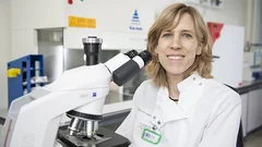
Health Innovation
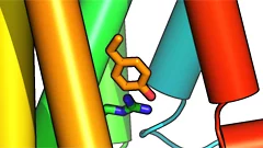
In the area of Health Innovation, several research groups at PSI are engaged in the study of fundamental questions regarding biology and cancer therapy. For example, they explore the structure of proteins – extremely complex biomolecules that are responsible for many different functions in organisms. Using PSI’s large research facilities, scientists also explore processes in biological tissue in order to fully understand their function and the development of specific diseases or deterioration processes. The ultimate goal is to find medicines that allow people to live as healthy a life as possible.
Patients with specific types of cancer are treated at the proton therapy facility on the PSI campus. Radiopharmaceuticals provide cancer treatments for very small tumours distributed throughout the body.
Find out more at: Overview Health Innovation
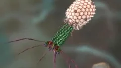
Hitting cancer from the inside
Researchers at the Paul Scherrer Institute PSI are now investigating a new method to channel radioactive substances directly into the nucleus of a cancer cell. Through this approach, the radiation source remains inside the cell and works in a more targeted way, because it gets closer to the cell's genetic information.

Medicines made to order with pinpoint precision
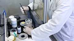
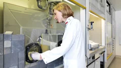
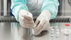
At PSI, scientists are developing new medicines against cancer. These contain radioactive substances that can be injected into the patients and thus make their way to the tumour. There, in direct contact, their radiation should destroy the cancer cells. Before such a radioactive medicine can be tested on patients in the first clinical trials, however, its safety must be guaranteed to ensure that the patient will not be harmed. Therefore every agent is produced at the PSI under sterile conditions and tested – separately for each patient, and only on the doctor's order.
Developing a new drug against thyroid cancer
Researchers at the Paul Scherrer Institute PSI have developed a drug to trace and treat a particularly malignant strain of thyroid cancer more effectively. One advantage of the new drug is that it can be used to treat a strain of thyroid cancer where the established treatment is ineffective. The researchers at PSI have developed the new drug to such an extent that an initial study conducted on cancer patients at the University Hospital Basel can now get underway.
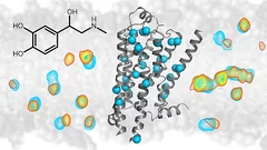
Probing what sets the heart racing
New insights into the workings of important drug receptorsMany medical drugs operate on specific receptors located in the outer walls of our body’s cells. One of these is called the beta-1 adrenergic receptor. Among other things, it is responsible for palpitation, the racing pulse that we feel with stage fright or infatuation. How it transmits signals to the cellular interior can now be revealed in detail. These findings could help scientists better understand many drugs' mode of action.
Targeting cancer
There are tumours where nothing seems to help: not chemotherapy, not external radiation therapy, not an operation. Often, they have already metastasised and can no longer be destroyed using conventional methods. The only option left here is internal radiotherapy with targeted radioactive drugs that strike directly at the heart of the disease. In order to make this possible, twenty specialists have been conducting research at the Centre for Radiopharmaceutical Sciences at the Paul Scherrer Institute PSI, a joint facility of PSI, ETH Zurich and the University Hospital Zurich.
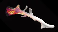
3D nanostructure of a bone made visible
Bones are made up of tiny fibres that are roughly a thousand times finer than a human hair. Researchers at the Paul Scherrer Institute PSI have developed a new computer-based algorithm with which they were able to visualize the localised order and alignment of these nanostructures inside an entire piece of bone for the first time.
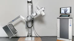
Robust X-ray machine for developing countries
The Paul Scherrer Institute PSI is involved in a project conducted by several research institutes (spearheaded by EPFL) to devise an X-ray machine especially for developing countries. The device should be able to cope with tropical climes and be easy and cheap to repair. PSI researchers are focusing on producing a cost-effective detector that is necessary for the imaging. The detector registers the X-ray light much like a chip in a digital camera.
New details of the transmission of stimuli in living organisms unveiled
Researchers unveil new details of how cells in a living organism process stimuli. So-called G-proteins, which help conduct external stimuli that reach a cell into its interior, play a central role here. For the first time, the study shows which parts of the G-proteins are vital for their function. Researchers from the Paul Scherrer Institute PSI, ETH Zurich, the pharmaceutical company Roche and the British MRC Laboratory of Molecular Biology report their results in the journals Nature and Nature Structural and Molecular Biology.

Fighting tumours with protons
Interview with Damien Charles WeberDamien Charles Weber has been the head and chief physician of the Centre for Proton Therapy, the only centre of its kind in Switzerland, since 2013. In this interview, he talks about the successes of proton therapy in cancer treatment and the objectives for the next few years in this field.
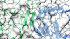
Together, not alone
Decoding biomolecules at SwissFEL and SLSProteins are a coveted but stubborn research object. A method developed for x-ray free-electron lasers and PSI’s future SwissFEL should now help researchers to make good headway in this field. It involves x-raying many small, identical protein samples consecutively at short intervals, thereby avoiding the main problem that protein research has faced thus far: producing samples in a sufficient size.

Phase contrast improves mammography
Phase contrast X-ray imaging has enabled researchers at ETH Zurich, the Paul Scherrer Institute (PSI) and the Kantonsspital Baden to perform mammographic imaging that allows greater precision in the assessment of breast cancer and its precursors. The technique could improve biopsy diagnostics and follow-up.
An infection tool with a metallic core
Thanks to the analysis of protein samples at the PSI, Lausanne researchers have managed to demonstrate which instrument bacteria use to transmit diseasesResearchers from ETH Lausanne EPFL have described how a particular strain of bacteria transmits diseases with unprecedented precision. The team of scientists headed by Petr Leiman, an assistant professor at the EPFL’s Laboratory of Structural Biology and Biophysics, demonstrated that the tip of a bacterial infection tool consists of a PAAR protein, which envelops a metal atom and tapers off to a sharp point. The findings are based on measurements carried out at the Swiss Light Source (SLS), one of the three large research facilities at the Paul Scherrer Institute (PSI).
How botox binds to neurons
Botox is a highly dangerous toxin that causes paralysis. In cosmetic applications it is used to temporarily eliminate wrinkles and in medicine as a treatment for migraine or to correct strabismus. An international research team has now established how the toxin molecule binds to the neuron whose activity is then blocked by the poison. The findings may be useful for the development of improved drugs with a lower risk of overdosage.
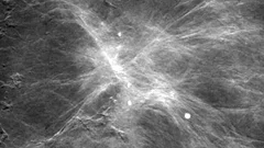
A promising new method for the diagnosis of breast cancer
A new mammography procedure that could generate substantial added value for the diagnosis of breast cancer in medical practice has just been published in the scientific journal Investigative Radiology. The method was developed at PSI in cooperation with the Certified Breast Centre at the Kantonsspital (cantonal hospital in) Baden and Philips as an industrial partner and is making the tiniest tissue changes visible. This has the potential to improve the early detection of breast cancer. Further studies in women suffering from breast cancer are to prove in a definitive manner the added value of the method.
Vitamins join fight against cancer
Cristina Müller, from the Center of Radiopharmaceutical Sciences at Paul Scherrer Institute (PSI), is researching a cancer therapy with radioactively labelled folate compounds. These enter the tumour cell unimpeded like a Trojan horse which is then killed as a consequence of emitted particle-radiation she explains.
Pancreas: new procedure detects tumours more efficiently
Better than CT and MRI: researchers at the Inselspital Berne, the University Hospital Basel and the Paul Scherrer Institute have devised a new method to detect small tumours in the pancreas.

A glimpse inside the control centres of cell communication
Numerous processes taking place within our body, such as sight, smell or taste, are accomplished by an important family of sensors on cell surfaces, which are known as G protein-coupled receptors (GPCR). Researchers have now compared the hitherto known structures of GPCRs and discovered a stabilising framework of fine struts that is characteristic for the architecture of the entire GPCR family. Knowledge about this constructional feature, which has been conserved over the course of evolution, can be of significant assistance in the development of new pharmaceuticals.
How stabilised cell fibres prevent cancer cell division
Anti-cancer drugs are used under the heading of Chemotherapeutics to prevent cells from dividing. Because the cells in a growing tumour divide more frequently than others, tumour cells are damaged more severely. Scientists at the Paul Scherrer Institute and the ETH Zurich have now clarified the exact mechanism of action of one class of these drugs. The data acquired is so accurate, that targeted drugs could now be developed that are even better suited to fulfil their task.
Alzheimer plaques in 3D
Researchers have succeeded in generating detailed three-dimensional images of the spatial distribution of amyloid plaques in the brains of mice afflicted with Alzheimer’s disease. The new technique used in the investigations provides an extremely precise research tool for a better understanding of the disease. In the future, scientists hope that it will also provide the basis for a new and reliable diagnosis method. The results were achieved within a joint project of two research teams à one from the Paul Scherrer Institute (PSI) and ETH Zurich, the other from the École Polytechnique Fédérale de Lausanne (EPFL).
Wenn die Datenleitung in die Zelle versagt
Lebende Zellen empfangen dauernd Informationen von aussen, die über Rezeptoren in das Zellinnere weitergeleitet werden. Genetisch bedingte Fehler in solchen Rezeptoren sind der Grund für zahlreiche Erbkrankheiten darunter verschiedene hormonelle Funktionsstörungen oder Nachtblindheit. Forschern des Paul Scherrer Instituts ist es nun erstmals gelungen, die exakte Struktur eines solchen fehlerhaften Rezeptors aufzuklären.This news release is only available in German.
