
Lab News & Scientific Highlights
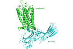
New insight into receptor signalling
A team of 72 investigators across 25 institutions including researchers from the Paul Scherrer Institut obtained the X-ray structure of a rhodopsinàarrestin complex, which represents a major milestone in the area of G-protein-coupled-receptor (GPCR), a protein family recognized in the award of the 2012 Nobel Prize in Chemistry.
Element-Specific X-Ray Phase Tomography of 3D Structures at the Nanoscale
Recent advances in fabrication techniques to create mesoscopic 3D structures have led to significant developments in a variety of fields including biology, photonics, and magnetism. Further progress in these areas benefits from their full quantitative and structural characterization.
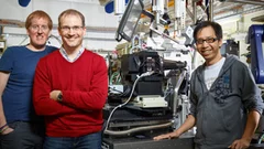
Together, not alone
Decoding biomolecules at SwissFEL and SLSProteins are a coveted but stubborn research object. A method developed for x-ray free-electron lasers and PSI’s future SwissFEL should now help researchers to make good headway in this field. It involves x-raying many small, identical protein samples consecutively at short intervals, thereby avoiding the main problem that protein research has faced thus far: producing samples in a sufficient size.
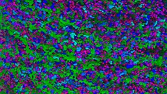
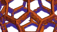
From inside an eggshell
Tiny cavities inside eggshells supply the materials that stimulate and control the shell’s growth. Using a novel imaging technique, researchers from the Paul Scherrer Institute (PSI), ETH Zurich and the Dutch FOM Institute AMOLF have succeeded in depicting these voids in 3D for the first time. In doing so, they lift an old limitation of tomographic images and hope that one day medicine will also benefit from their method.
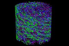
Multiresolution X-ray tomography, getting a clear view of the interior
Researchers at PSI have developed a technique that combines tomography measurements at different resolution levels to allow quantitative interpretation for nanoscale tomography on an interior region of interest of the sample. In collaboration with researchers of the institute AMOLF in the Netherlands and ETH Zurich in Switzerland they showcase their technique by studying the porous structure within a section of an avian eggshell. The detailed measurements of the interior of the sample allowed the researchers to quantify the ordering and distribution of an intricate network of pores within the shell.
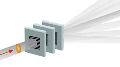
Split x-ray flash shows rapid processes
SwissFEL, PSI’s x-ray laser, is to render the individual steps of very rapid processes visible. A new method will facilitate especially precise experiments: the individual x-ray flashes are split into several parts that arrive at the object under examination one by one. The principle of the method harks back to the ideas of the earliest high-speed photography.
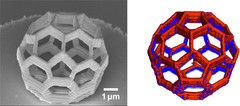
Nanometres in 3D
Scientists at the Paul Scherrer Institute and ETH Zurich have created 3D images of tiny objects showing details down to 25 nanometres. In addition to the shape, the scientists determined how particular chemical elements were distributed in their sample and whether these elements were in a chemical compound or in their pure state.
Shortcut to protein portraits
All living organisms, from bacteria to humans, rely on proteins to perform their vital functions. How these proteins accomplish their tasks depends on their structure. Researchers from the Paul Scherrer Institute have now devised a novel method to determine the crystal structure of proteins using X-ray light, which could also hasten the development of new drugs in future. The study will be published in the journal Nature Methods on 15 December.
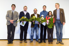
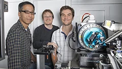
Innovation Award on Synchrotron Radiation 2014 for high-resolution 3D hard X-ray microscopy
The 2014 Innovation Award on Synchrotron Radiation was bestowed to researchers Ana Diaz, Manuel Guizar-Sicairos, Mirko Holler, and Jörg Raabe from the Paul Scherrer Institut, Switzerland, for their contributions to method and instrumentation development, which have set new standards in high-resolution 3D hard X-ray microscopy.