
Scientific Highlights and News

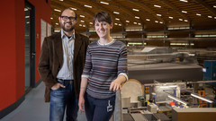
Dr. Manuel Guizar-Sicairos elected as Fellow member of The Optical Society (OSA)
Dr. Manuel Guizar-Sicairos, beamline scientist at the cSAXS beamline, was elected as a Fellow Member of The Optical Society (OSA) for seminal contributions to methods and applications of coherent lensless imaging, ptychography, x-ray nanotomography, and new modalities of x-ray microscopy.
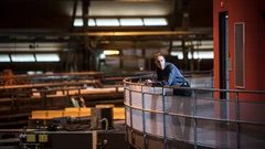
Nanoworlds in 3-D
Tomographic images from the interior of fossils, brain cells, or computer chips are yielding new insights into the finest of structures. These 3-D images are made possible by the X-ray beams of the Swiss Light Source SLS, together with detectors and sophisticated computer algorithms developed at PSI.
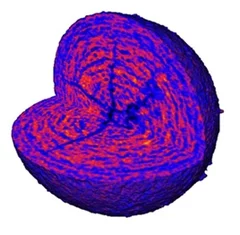
Short film of a magnetic nano-vortex
Using a newly developed imaging method, researchers were able to visualise the magnetic structure inside a material with nanoscale resolution. They succeeded in creating a short "film" consisting of seven movie frames that shows, for the first time in 3D, how tiny vortices of the magnetisation deep within a material change over time.
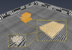
3D imaging for planar samples with zooming
Researchers of the Paul Scherrer Institut have previously generated 3-D images of a commercially available computer chip. This was achieved using a high-resolution tomography method. Now they extended their imaging approach to a so-called laminography geometry to remove the requirement of preparing isolated samples, also enabling imaging at various magnification. For ptychographic X-ray laminography (PyXL) a new instrument was developed and built, and new data reconstruction algorithms were implemented to align the projections and reconstruct a 3D dataset. The new capabilities were demonstrated by imaging a 16 nm FinFET integrated circuit at 18.9 nm 3D resolution at the Swiss Light Source. The results are reported in the latest edition of the journal Nature Electronics. The imaging technique is not limited to integrated circuits, but can be used for high-resolution 3D imaging of flat extended samples. Thus the researchers start now to exploit other areas of science ranging from biology to magnetism.
Inside Batteries
Lithium ion batteries (LIB) are essential in modern everyday life, with increasing interest in enhancing their performance and lifetime. Secondary particles of Li-rich cathode material were examined with correlated ptychographic X-ray tomography and diffraction microscopy at different stages of cycling to probe the aging mechanism.
Virtual lens improves X-ray microscopy
A method developed by PSI researchers makes X-ray images of materials even better. The researchers took a number of individual images while moving an optical lens. From these, with the help of computer algorithms, they generated one overall image.