
News & Scientific Highlights
Making the world go round - a look into the structure of a prominent heterogeneous catalyst
Fluid catalytic cracking catalysts, which are composite particles of hierarchical porosity, were examined using ptychographic X-ray tomography. These particles are essential to the conversion of crude oil into gasoline. Examination of catalysts at decreasing levels of catalytic conversion efficacy allowed the detection of possible deactivation causes.
Swiss Neutron Scattering Prize
Viviane Lutz-Bueno was awarded the Swiss Neutron Scattering Prize of the Swiss Neutron Scattering Society, at the Joint Annual Meeting of the Swiss and Austrian Physical Society, for her PhD thesis Effects of formulation and flow on the structure of micellar aggregates carried out at ETH Zürich. Viviane is currently a postdoctoral fellow at the CXS group at PSI, developing scanning SAXS analysis methods.
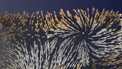
Diving into magnets
For the first time, scientists have made visible the directions of the magnetisation inside a 3D magnetic object. The smallest details in their visualisation were ten thousand times smaller than a millimetre. Among others, the magnetic structure contained one outstanding kind of pattern: magnetic singularities called Bloch points, which up to now were only known in theory.
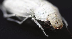
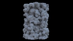
Photonic structure of white beetle wing scales: optimized by evolution
A very thin layer on this beetle’s wings exhibits a complicated structure on the nanoscale that gives them a bright white color. X-ray nanotomography acquired at the Swiss Light Source provides a faithful image of this structure in three dimensions with which scientists can confirm its evolutionary optimization: just enough material for an efficient reflection of white light.
European NESY Winterschool Young Scientist Best Poster Prize
Klaus Wakonig was awarded the "Young Scientist Best Poster Prize" along with a cash prize at the 10th European NESY Winterschool & Symposium on Neutron and Synchrotron Radiation. Klaus is a PhD student at the coherent X-ray scattering group (CXS) in PSI. His poster, entitled "X-ray Fourier ptychography," details his latest results in the implementation of Fourier ptychography at X-ray wavelengths for nanoimaging. Image credit ©NESY/Montanuniversitaet Leoben
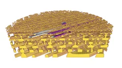
3-D X-ray imaging makes the finest details of a computer chip visible
Researchers at the PSI have made detailed 3-D X-ray images of a commercially available computer chip. In their experiment, they examined a small piece that they had cut out of the chip beforehand. This sample remained undamaged throughout the measurement. It is a major challenge for manufacturers to determine if, in the end, the structure of their chips conforms to the specifications. Thus these results represent one important application of an X-ray tomography method that the PSI researchers have been developing for several years.
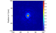
Interlaced zone plates push the resolution limit in x-ray microscopy
A novel type of diffractive lenses based on interlaced structures enable x-ray imaging at resolutions below 10 nm. The fabrication method and the test results of these novel x-ray lenses have been published in the journal Scientific Reports.
How does food look like on the nanoscale?
The answer to this question could save food industry a lot of money and reduce food waste caused by faulty production. Researchers from the University of Copenhagen and the Paul Scherrer Institut have obtained a 3D image of food on the nanoscale using ptychographic X-ray computed tomography. This work paves the way towards a more detailed knowledge of the structure of complex food systems.
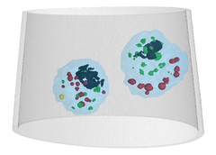
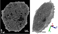
Mass density distribution of intact cell ultrastructure
The determination of the mass density of cellular compartments is one of the many analytical tools that biologists need to unravel the extremely complex structure of biological systems. Cryo X-ray nanotomography reveals absolute mass density maps of frozen hydrated cells in three dimensions.