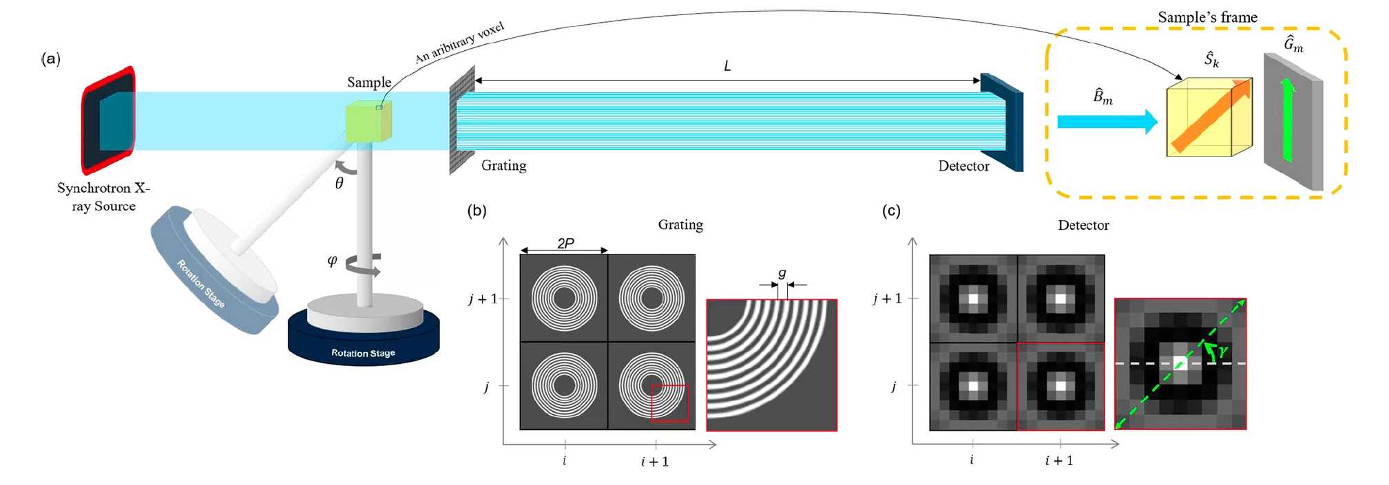
X-ray grating interferometry provides three complementary signals, based on absorption, differential phase and small-angle scattering contrast. X-ray scattering imaging can give access to microstructural information for features well below the setup resolution, in a large field of view, making this technique very interesting for the investigation of new materials. Recently, 2D omni-directional X-ray scattering sensitivity in a single shot has been demonstrated, paving the way to time resolved studies. The objective of this study is to extend 2D omni-directional X-ray scattering imaging to 3D without need for a priori knowledge on the scatterer shape and/or space organization. The proposed project will provide a more generalized directional sensitivity than achieved so far without need for a-priori knowledge on the scatterer shape and/or space organization. This aspect is important to efficiently study samples with structures of unknown orientation or with features with an orientation varying in space and time. The single shot characteristic of this technique will promote in-situ and/or time resolved studies at synchrotron facilities and open-up new opportunities for cutting-edge materials science applications.
Publications
- Kim J, Kagias M, Marone F, Stampanoni M, X-ray scattering tensor tomography with circular gratings,
Applied Physics Letters. 2020; 116(13): 134102 (5 pp.). - Kim J, Kagias M, Marone F, Shi Z, Stampanoni M, Fast acquisition protocol for X-ray scattering tensor tomography, Scientific Reports. 2021; 11(1): 23046 (13 pp.) 2021.
- Kim J, Slyamov A, Lauridsen E, Birkbak M, Ramos T, Marone F, et al., Macroscopic mapping of microscale fibers in freeform injection molded fiber-reinforced composites using X-ray scattering tensor tomography, Composites Part B: Engineering. 2022; 233: 109634 (11 pp.).
Collaboration
- Computational Imaging group, CWI, The Netherlands
- Composite group, Chalmers University, Sweden
- Xnovo Technology ApS, Denmark
Funding
- European Union’s Horizon 2020 research and innovation programme (Marie Skłodowska-Curie Grant Agreement No. 765604)
- EUROSTARS INFORMAT E! 11060 project
