In this project, we aim to investigate, promote and facilitate the use of X-ray phase contrast microtomography as a complementary method for histopathological techniques in the clinical environment.
Examination and visualization of biological tissues in Pathology is the essential diagnostic method in medical practice. Currently used histological techniques rely on chemical fixation, staining, very thin sectioning and subsequent visible microscopy inspection of the tissue specimens extracted from the patient’s body.
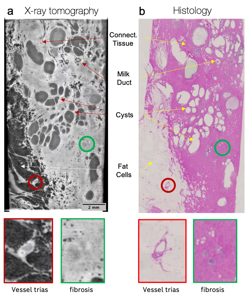
Exploiting the higher sensitivity of X-ray phase contrast is particularly suited for biological soft tissues. Therefore, the proposed approach may substantially reduce the staining requirements of the specimens, thus allowing their examination in conditions closer to their natural state. In addition, X-ray microtomography does not destroy the tissue as the sectioning is virtually done a posteriori on its three-dimensional tomographic image reconstruction.
In our team, a prototype of the new X-ray uCT setup was assembled. Based on the grating interferometry, the system provides high spatial resolution (<10 µm) and field of view (up to 2.0 cm). Therefore, the three-dimensional information obtained from X-ray phase-contrast microtomography nicely corresponds to the images obtained by conventional histological techniques.
The proposed high-resolution CT system for soft-tissue imaging closes the gap between Radiology and Pathology. The main focus of our current research is developing a prototype of the X-ray uCT system with clinically compatible image quality and acquisition time.
Publications
- T. Thüring, P. Modregger, T. Grund, J. Kenntner, C. David, and M. Stampanoni, High resolution, large field of view x-ray differential phase contrast imaging on a compact setup. Applied Physics Letters, 99, 041111 (2011)
- Vila-Comamala, J. et al. High sensitivity X-ray phase contrast imaging by laboratory grating-based interferometry at high Talbot order geometry. Optics Express 29, 2049 (2021).
Collaboration
- Prof. Dr. med. Gad Singer, Chefarzt, Institute of Patology, Kantonsspital Baden AG (Switzerland)
- Prof. Dr.med Zsuzsanna Varga, Leitende Ärztin Institut für Pathologie und Molekularpathologie, Koordinatorin Brustzentrum
Funding
- FNSNF Grant Number 159263
- ERC Grant ERC-2012-StG 310005-PhaseX
Contacts
- Dr. Maxim Polikarpov, maxim.polikarpov@psi.ch