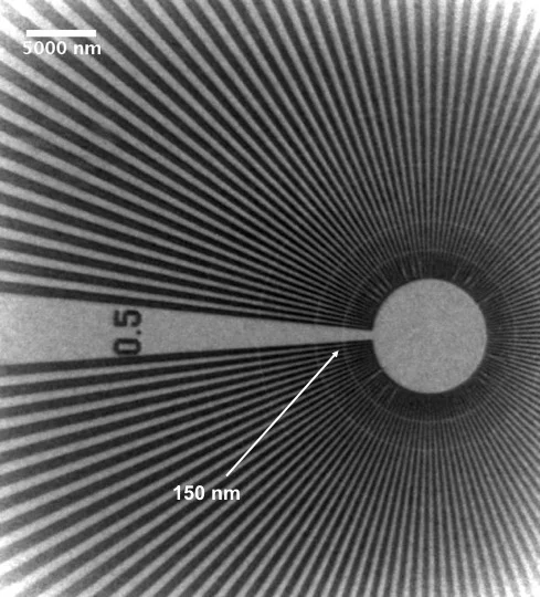
At modern third generation synchrotron sources, voxel sizes in the micrometer range are routinely achieved. However, isotropic 100 nm barrier is reached and surpassed by only a few instruments. At the TOMCAT beamline of the Swiss Light Source, the multimodal endstation (which offers tomographic capabilities in the micron range) is equipped with a full field, hard X-ray nanoscope.
This full-field transmission X-ray microscope (TXM) is composed of custom optical components. A condenser (a custom designed beamshaper) produces a top-flat illumination in the focal plane. After the sample, the Fresnel Zone Plate (FZP), with a focal length chosen according to the energy, acts as an objective lens. To exploit phase contrast, phase rings can be inserted in the back-focal distance of the FZP to generate positive or negative Zernike phase contrast. The detector, placed downstream at about 10 m, records the magnified image of the sample.
The latest improvements in optical and detector designs as well as stability of the hardware lead to a field-of-view from 50 to 80 μm2 with a pixel size down to 60nm in the 8-20 keV energy range.
In the case of the broadband radiation use, tomographic investigations can be performed within few minutes. The setup can be used in absorption or phase contrast mode for a wide range of applications, including biology, geology, materials science and paleontology.
The ongoing efforts aim to further improve the hard components (optics and setup versatility) as well as data acquisition speed, spatial resolution and sensitivity.
Publications
- Stampanoni M. et al. "Phase-contrast tomography at the nanoscale using hard x rays." Physical Review B 81, 140105 (2010).
- Vartiainen I. et al. "Halo suppression in full-field x-ray Zernike phase contrast microscopy." Opt. Lett. 39, 1601 (2014).
Collaboration
Laboratory for Micro and Nanotechnology, Paul Scherrer Institut, Switzerland
Contact
