Aller au contenu principal
  • Paul Scherrer Institut PSI
  • PSI Research, Labs & User Services

Digital User Office

  • Digital User Office
  • DE
  • EN
  • FR
Paul Scherrer Institut (PSI)
Rechercher
Paul Scherrer Institut (PSI)

Hauptnavigation

  • Research at PSIOuvrir ce point de menu principal
    • Research Initiatives
    • Ethics and Research integrity
    • Scientific Highlights
    • Scientific Events
    • Scientific Career
    • PSI-FELLOW
    • PSI Data Policy
  • Research Divisions and LabsOuvrir ce point de menu principal
    • Overview
    • Research with Neutrons and Muons
    • Photon Science
    • Energy and Environment
    • Nuclear Energy and Safety
    • Biology and Chemistry
    • Scientific Computing, Theory and Data
    • Large Research Facilities
    • Logistics
  • Facilities and InstrumentsOuvrir ce point de menu principal
    • Overview
    • Large Research Facilities
    • Facilities
    • PSI Facility Newsletter
  • PSI User ServicesOuvrir ce point de menu principal
    • User Office
    • Methods at the PSI User Facilities
    • Proposals for beam time
    • Proposal Deadlines
    • Data Analysis Service (PSD)
    • EU support programmes
  • New ProjectsOuvrir ce point de menu principal
    • SLS 2.0
    • IMPACT
  • DE
  • EN
  • FR

Digital User Office (mobile)

  • Digital User Office

Vous êtes ici:

  1. PSI Home
  2. Labs & User Services
  3. PSD
  4. LSC
  5. MicroSpec Group
  6. Scientific Highlights and News
  7. Soft X-ray Laminography – a new 3D soft X-ray imaging technique

Navigation secondaire

MicroSpec

  • People
  • Research
  • Scientific Highlights and News
  • Pixelator
  • Publications

Info message

Ce contenu n'est pas disponible en français.
27 janvier 2020

Soft X-ray Laminography: 3D imaging with powerful contrast mechanisms

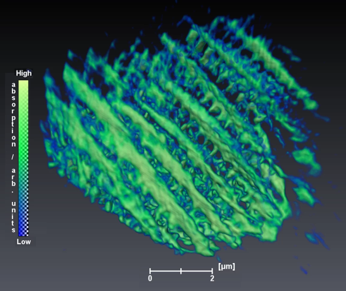
Inner Structure of a Butterfly Wing Scale
3D model constructed from soft X-ray laminography measurements of the front tip of the wing scale from a European peacock butterfly.

Soft X-ray 3D imaging has already been realized at synchrotron radiation sources using either scanning transmission X-ray microscopy (STXM) schemes or tomography-based concepts. However, the maximum accessible sample volume is severely limited by the reduced penetration depth of the lower-energy soft X-ray radiation. This becomes even more of a drawback in the case of flat and extended specimens, which can be found in various fields of nanoscience.

The generalized geometry of laminography, characterized by a tilted axis of rotation concerning the incident X-ray beam resulting in a constant material thickness during rotation, has proven to be particularly suitable for the investigation of laterally extended and thin objects. The combination of soft X-rays and laminography provides the unique potential of bridging the gap between investigations of elaborate nanostructured thin film samples and taking advantage of the characteristic absorption contrast mechanisms in the soft X-ray range.

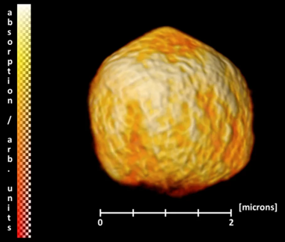
SPION coated microballoon
Three-dimensional image of an air-filled PVA microsphere coated with magnetite nanoparticle agglomerates reconstructed from soft X-ray laminography measurements.

In collaboration with scientists from the Friedrich-Alexander-Universität Erlangen-Nürnberg, the University of Cambridge and colleagues from the cSAXS beamline of the Swiss Light Source, the research team of the PolLux endstation of the Swiss Light Source developed the new 3D imaging technique of Soft X-ray Laminography (SoXL). While the PolLux STXM already provides high spatial (nm-range) and energy resolution, its extension towards SoXL allows for a detailed three-dimensional visualization and analysis of the inner structure of sophisticated, microstructured, and magnetic objects.

The scientists demonstrated the wide range of current research areas that can be investigated using SoXL, using various examples of applications from functionalized nanomaterials, biological nanostructures with photonic properties and sophisticated magnetic materials. Especially for the latter, the properties of soft X-rays played a decisive role, providing an enhanced magnetic contrast at the L absorption edges of transition metals.

SoXL is running as a standard operation tool at the PolLux beamline and is now available for user operation of the broad community of nanomaterials and nanoscience.

Contact:
Dr. Jörg Raabe
Swiss Light Source
Paul Scherrer Institut
Telephone: +41 56 310 5193
E-mail: joerg.raabe@psi.ch


Original Publication:
From 2D STXM to 3D Imaging: Soft X-ray Laminography of Thin Specimens
Katharina Witte, Andreas Sp
äth, Simone Finizio, Claire Donnelly, Benjamin Watts, Blagoj Sarafimov, Michal Odstrcil, Manuel Guizar-Sicairos, Mirko Holler, Rainer H. Fink, Jörg Raabe
Nano Letters, 2020
DOI:
10.1021/acs.nanolett.9b04782
 

Sidebar

top

Pied de page

Paul Scherrer Institut

Forschungsstrasse 111
5232 Villigen PSI
Suisse

Téléphone: +41 56 310 21 11
Téléfax: +41 56 310 21 99

Comment nous trouver 
Contact

Centre de visiteurs psi forum
Laboratoire élèves iLab (en allemand)
Centre de protonthérapie
Centre de Formation du PSI
PSI Guest House (en anglais)
PSI Gastronomie (en allemand)
psi forum shop

Service & Support

  • Annuaire
  • User Office
  • Accelerator Status
  • Publications du PSI
  • Fournisseurs
  • E-facture
  • Computing (en anglais)
  • Sicherheit (en allemand)

Carrière

  • Travailler au PSI
  • Offres d'emploi
  • Formation initiale et formation continue
  • Career Center
  • Formation professionnelle (en allemand)
  • Centre de Formation du PSI

Pour les médias

  • Le PSI en bref
  • Chiffres et faits
  • Le coin médias
  • Communiqués de presse
  • Réseaux sociaux

Suivez le PSI: Twitter (in English) LinkedIn Youtube Facebook Instagram Issuu RSS

Footer legal

  • Impressum
  • Conditions d'utilisation
  • Login éditeurs