

Research Interests
One of the major promises of X-ray free-electron laser (XFEL) technology is to advance structural biology from the determination of molecular snapshots to molecular movies. Together structural and dynamic information provides unique insights into the function of proteins as principal building blocks of our biology.
The key advantage of XFEL-based over conventional diffraction strategies stems from the characteristics of the XFEL pulses, which contain ~1012 X-ray photons but are only tens of femtoseconds long. This fluence is sufficient to produce high-resolution diffraction patterns from very small crystals, while at the same time outrunning most radiation damage processes. The ultrafast XFEL pulses further open up the possibility for time-resolved pump probe experiments on femtosecond to millisecond timescales. Together these advantages allow studying protein structural dynamics at unprecedented spatial and temporal resolution; essentially opening a new frontier in structural biology.
My group focuses on the implementation of time-resolved serial crystallography at the Swiss Light Source (SLS) and the Swiss Free Electron Laser (SwissFEL). Naturally light-sensitive retinal-binding proteins like bacteriorhodopsin are paving the way for time-resolved studies using XFELs. In the near future, we will further see the molecular details of how protons, chloride and sodium ions are pumped across biological membranes by light. My research further aims to answer the question of how protein interactions guide the high quantum efficiency and stereo selectivity of the ultrafast retinal isomerization. Time-resolved crystallography will help us understand fundamental biological processes including the high photo efficiency of the visual sense and how retinal proteins might be improved to better manipulate neural cells in the field of optogenetics.
Laser light allows unmatched precision which makes it an ideal trigger for time-resolved measurements. Learning from biology, chemists have developed a large repertoire of synthetic photoswitches with highly tunable properties. Like their natural counterpart retinal, these chromophores can be inserted into proteins to put them under optical control. In photopharmacological applications reversibly binding ligands are envisioned to precisely control pharmaceutical targets such as ion channels, GPCRs or microtubules. Compared to optogenetics the approach has a wider range of applications including medical intervention in humans since the method relies on the chemical manipulation of native proteins and is not dependent on genetic manipulation. Harnessing the chemistry of such synthetic photoswitches to study proteins that cannot be natively activated by light will dramatically increase the number of biological systems whose structural dynamics can be studied at modern XFEL sources. Dynamic information on how ligands influence a particular protein conformation will provide a crucial new dimension to molecular pharmacology.
Group Members

As a PI at PSI i am actively researching alternatives to light activation to study proteins by time-resolved crystallography. As a Senior Scientist in the Group of Jörg Standfuss i am the groups' expert in time-resolved serial crystallography.


News & Events
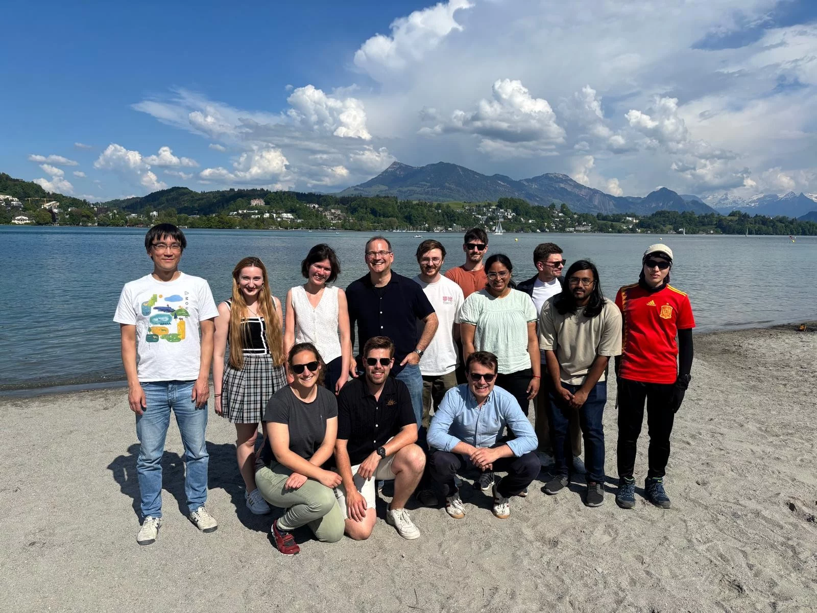
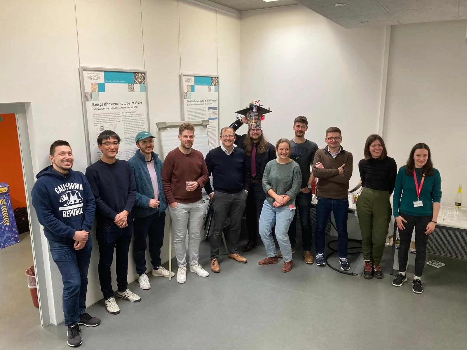
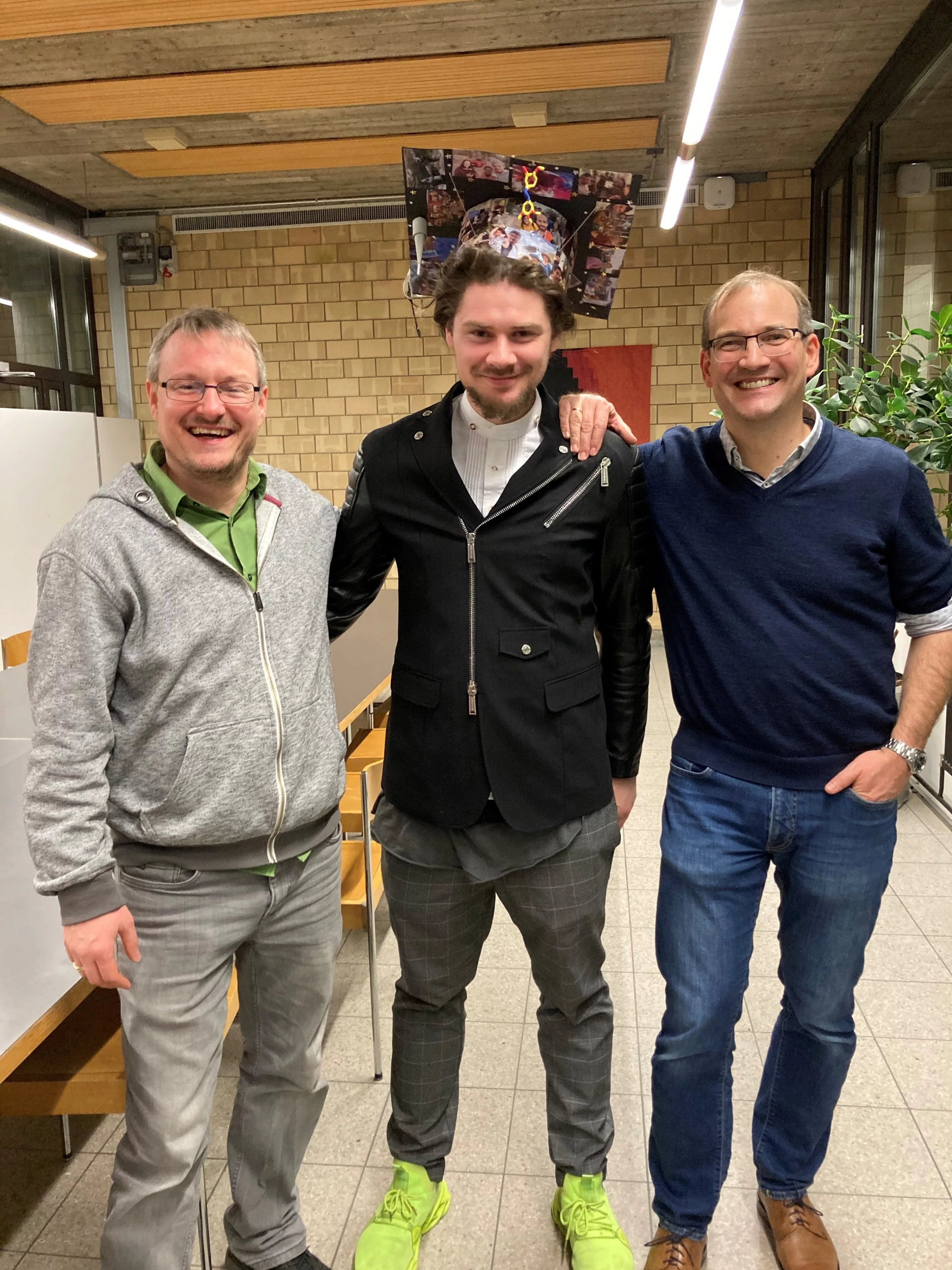
April 2025, Group Outing, Food Trail Luzern
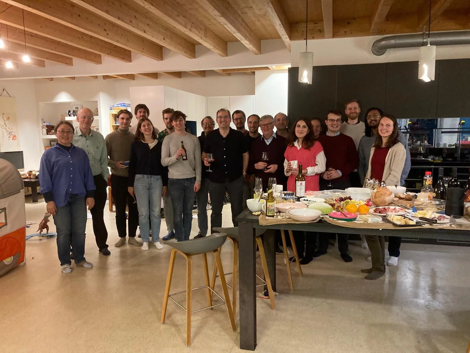
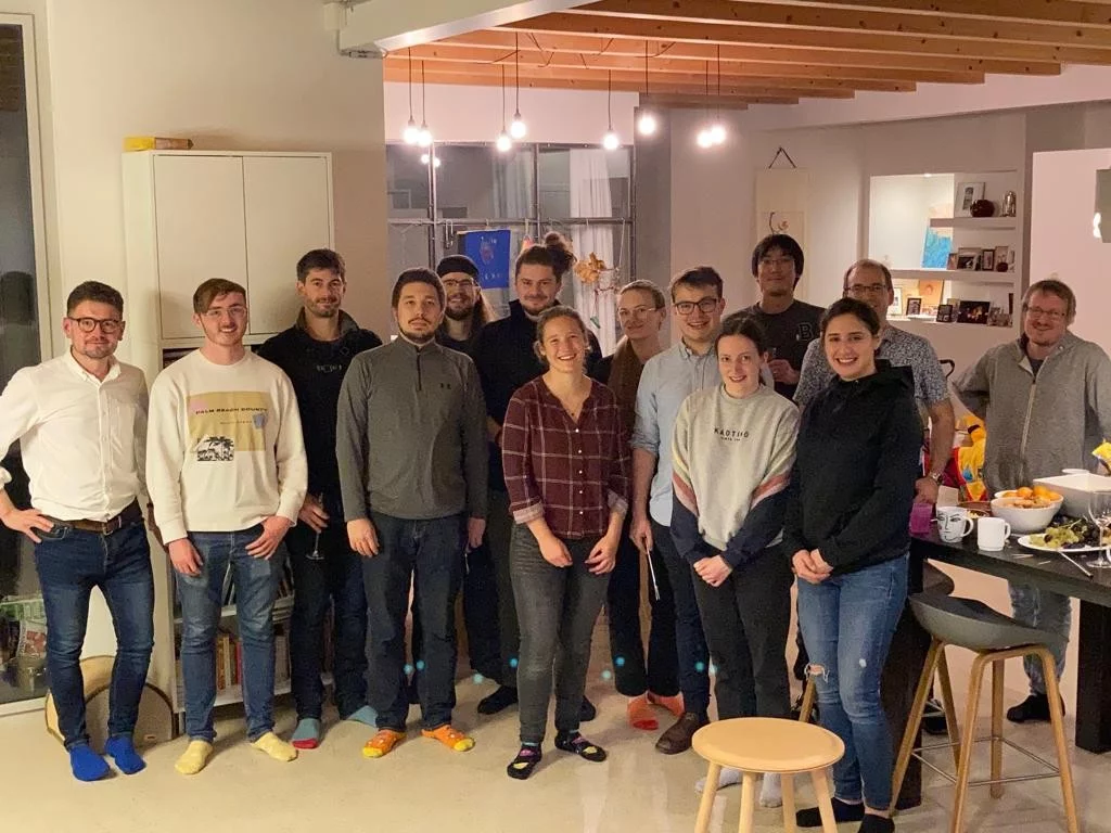
Dec 2024, Group Christmas Party
Dec 2024, PhD Thesis defense of Robin Stipp, congratulations Robin !!!
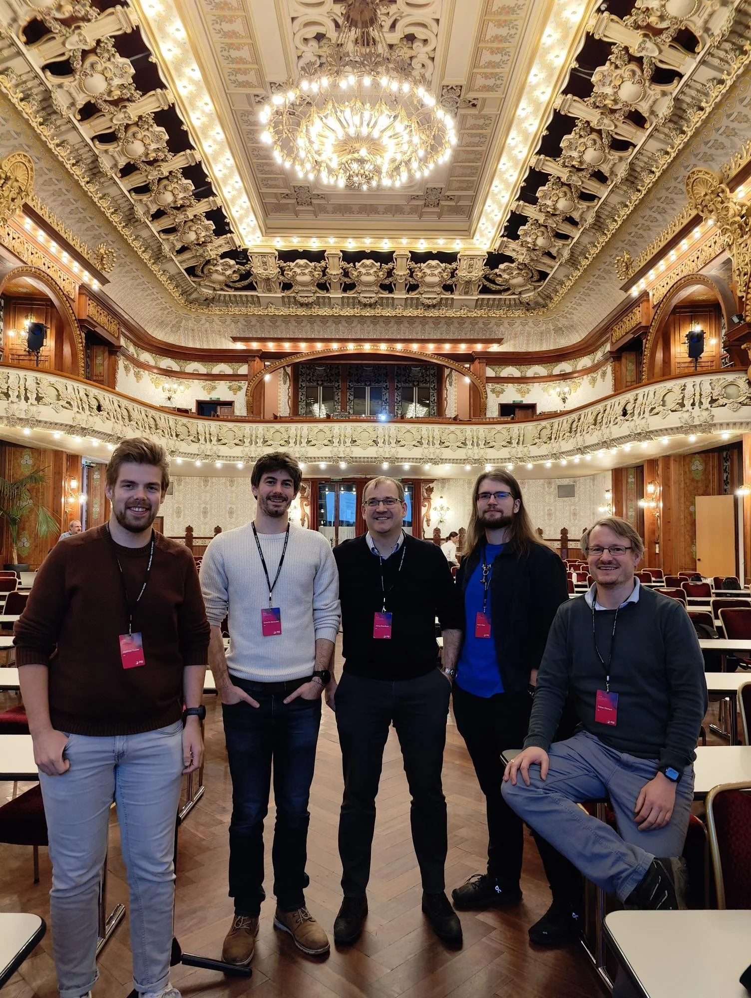
Nov 2024, International Conference on Retinal Proteins, Interlaken
May 2024, PhD Thesis defense of Melissa Carrillo, congratulations Melissa !!!

December 2023, PhD Thesis defense of Hannah Glover, congratulations Hannah !!!

September 2023, Group Hike Flumserberg

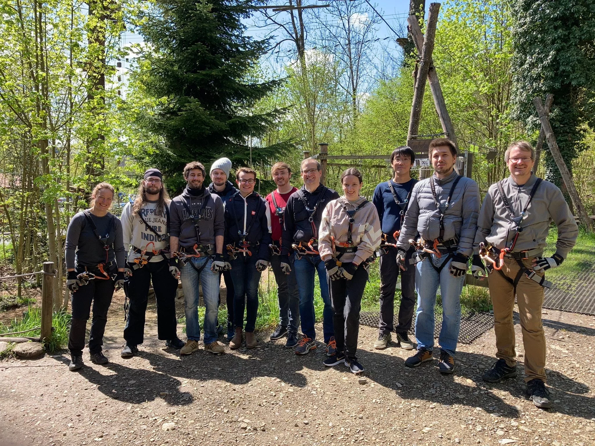
April 2023, Group Outing Adventure Park Rheinfall
04.03.2023, Der Kälte getrotz - Wissenschaftsmagazin - SRF
https://www.srf.ch/audio/wissenschaftsmagazin/der-kaelte-getrotzt?id=12342967
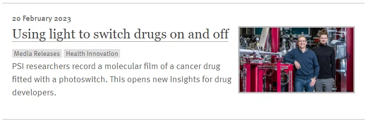
(18:10) Medikamente per Lichtimpuls ein- und ausschalten, dieses Forschungsfeld steckt noch in den Kinderschuhen. Am Paul Scherrer Institut PSI haben Forschende nun einen solchen lichtsensitiven Wirkstoff erstmals in Aktion gefilmt. Allein das schon ist besonders. Und: diese Filmsequenzen könnten der Medikamentenentwicklung insgesamt helfen.
Feb 2023, PSI Press Release "Using light to switch drugs on and off"
https://www.psi.ch/en/media/our-research/using-light-to-switch-drugs-on-and-off
Jan 2023, PhD Thesis defense of Maximilian Wranik, congratulations Max!!!
Dec 2022, Group Christmas Party
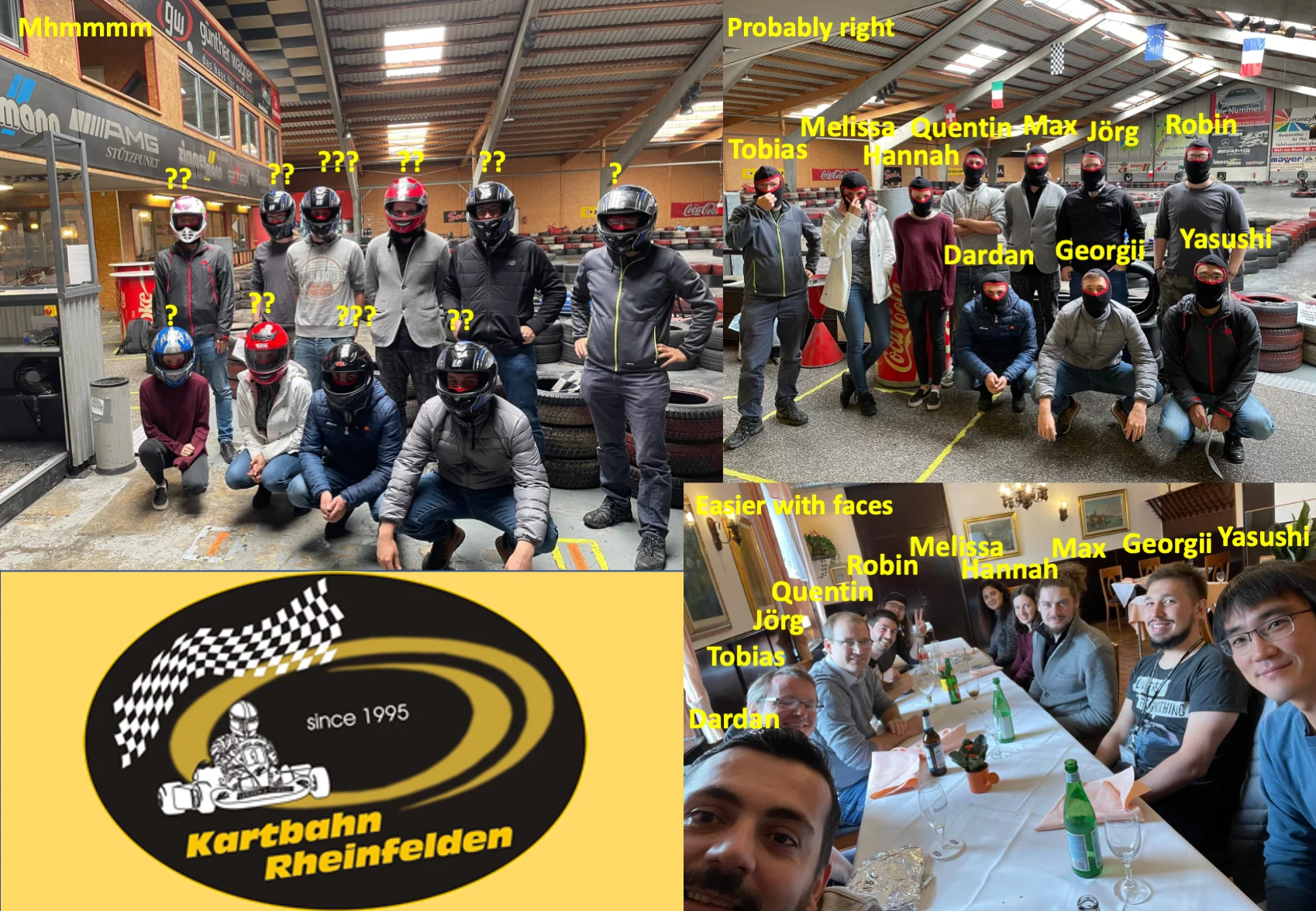
Oct 2021, Group Outing Rheinfelden
Jan 2021, PSI Thesis Medal 2021 for pioneering structural biology at SwissFEL, Congratulations Petr!!!!
https://www.psi.ch/en/bio/scientific-highlights/psi-thesis-medal-2021

Jul 2019, Muotatal and Hölloch Exploration

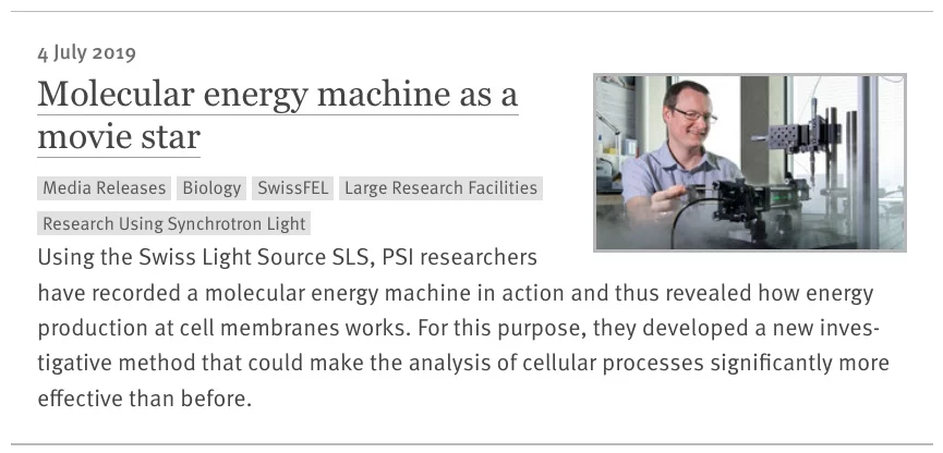
Jul 2019, Time-resolved crystallography now possible at the SLS
https://www.psi.ch/en/media/our-research/molecular-energy-machine-as-a-movie-star
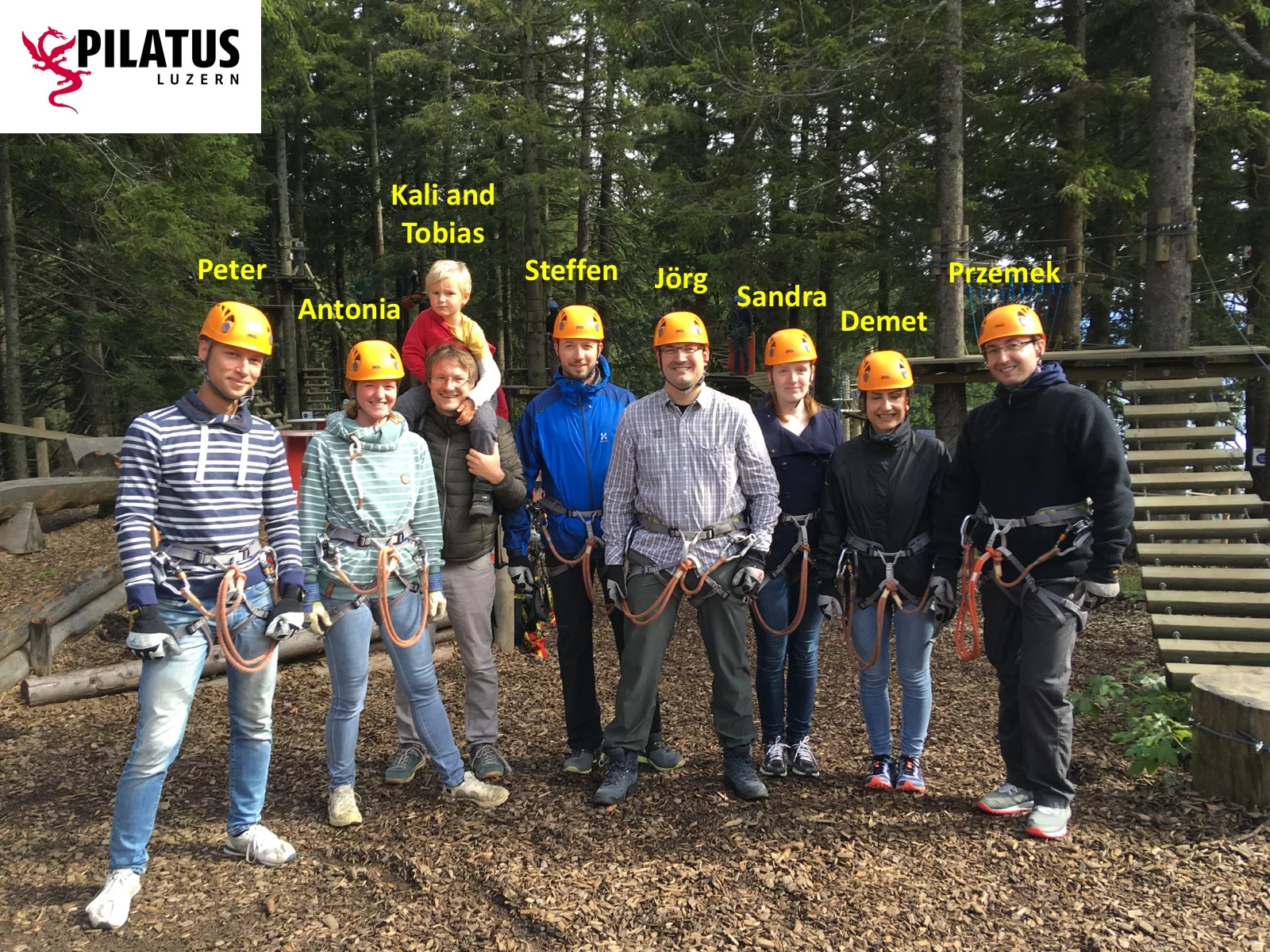
Oct 2018, Climbing at high wire park Pilatus
Jun 2018, Our research featured by the SLAC National Accelerator Laboratory
https://www6.slac.stanford.edu/news/2018-06-14-scientists-make-first-molecular-movie-one-nature%E2%80%99s-most-widely-used-light-sensors
Nov 2017, Przemek Nogly started group at the ETH Zurich on a SNSF Ambizione grant. Congratulations well deserved!!

Sep 2017, Demet Kekilli and Steffen Brünle joined the PSI-FELLOW-II programme co-funded by Horizon2020 of the European Commission

Jan 2017, Our research featured by the Swiss National Science Foundation
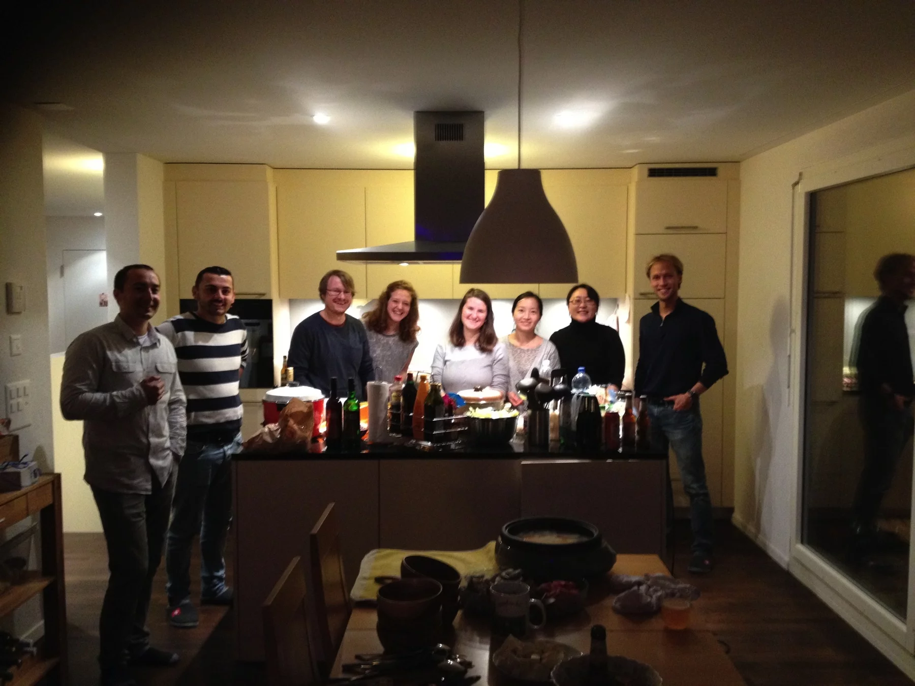
Dec 2016, Chäsfondue and Hotpot Event
Aug 2016, PSI Press Release "Catching Proteins in the Act"
https://www.psi.ch/media/catching-proteins-in-the-act
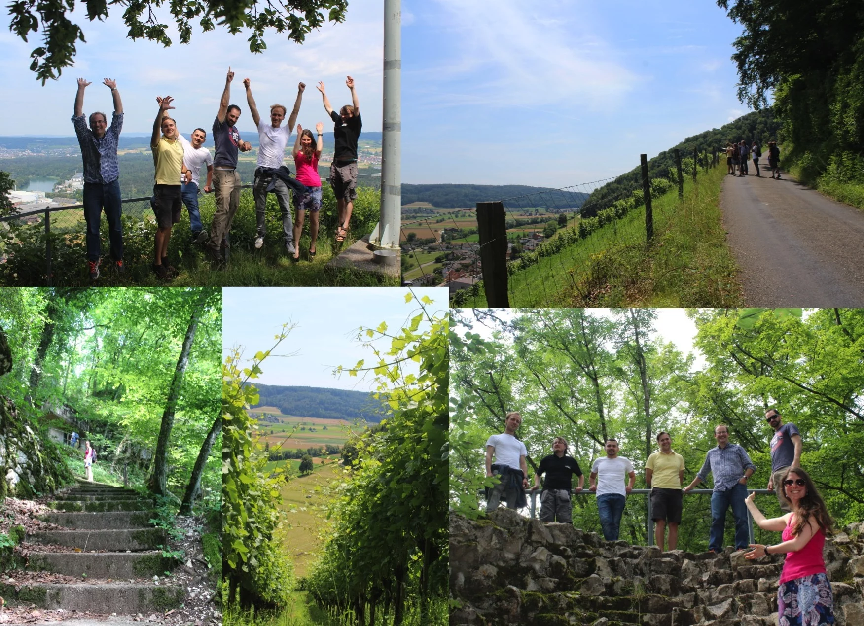
Jul 2016, Besserstein Hike
Former Members
Khusainov, Gerorgii
Ph.D. Student
Stipp, Robin
Ph.D. Student
Carrillo, Melissa
Ph.D. Student
Glover, Hannah
Ph.D. Student
Wranik, Maximilian
Ph.D. Student
James, Daniel Dr.
Scientist
Skopintsev, Petr
Ph.D. Student
Bünle, Steffen Dr.
Postdoc
Kekilli, Demet Dr.
Postdoc
Jaeger,Kathrin
Ph.D. Student
Singhal,Ankita
Ph.D. Student
Ostermaier, Martin
Ph.D. Student
Publications since 2010
Feb 2023, PSI Press Release "Using light to switch drugs on and off"
https://www.psi.ch/en/media/our-research/using-light-to-switch-drugs-on-and-off