
News & Scientific Highlights
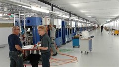
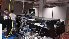
Last undulator placed in SwissFEL tunnel
On the 6th of October the last undulator for the ARAMIS beamline was placed into the SwissFEL tunnel. Thanks to the efficiency and motivation of the different groups involved with undulator preparation, all 12 undulators were assembled, measured and installed in the tunnel between the 2nd of February 2016 and the 6th of October 2016.
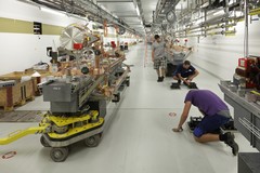
Completion of SwissFEL LINAC
On September 13th, the last two modules of the linear accelerator were installed in the SwissFEL tunnel. This means that 26 accelerating modules are installed now. One accelerating module consists of four accelerating structures. In total there are 104 accelerating structures, with a lenght of 2 m each.
SwissFEL First Free Electrons, First Beam at 144 MeV and First acceleration with SwissFEL C-band modules
For the first time electrons were accelerated with a SwissFEL C-band module (the first one of a series of 26 modules). The module operated with the nominal parameters that will be used in the last two linac sections. The RF pulse duration was 3 µs, at an RF power of 36 MW from the klystron. This pulse was compressed to 350 ns, yielding a peak power of approximately 215 MW. At these conditions, the energy gain was estimated to be 235 MeV, which is well within expectations.
First Free Electrons at SwissFEL
At SwissFEL the first free electrons were produced and accelerated to 7.9 MeV. The electrons were stopped directly after the gun in the gun-spectrometer. The bunch charge was 20-50pC, with a repition rate of 10Hz. First measurements showed that the generated electron beam was of high quality. This means that the first milestone for the SwissFEL beam commissioning was reached!
Catching proteins in the act
Proteins are indispensable building blocks of life. They play a vital role in many biological processes. Researchers have now been able to show how the ultrafast processes by which proteins do their work can be studied with free-electron X-ray lasers such as SwissFEL at the Paul Scherrer Institute PSI. Free-electron X-ray lasers generate extremely short and intense pulses of X-ray light. Currently there are just two such facilities in operation, worldwide. The results were published in the scientific journal Nature Communications.
Catching proteins in the act
Some of the fastest processes in our body run their course in proteins activated by light. The protein rhodopsin sees to it that our eyes can rapidly take in their ever-changing surroundings. Free-electron X-ray lasers such as SwissFEL at the Paul Scherrer Institute PSI now make it possible for the first time to catch such processes in flagranti. Free-electron X-ray lasers generate extremely short and intense pulses of X-ray light.
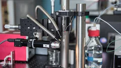
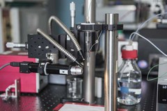
First light from the SwissFEL Experimental Laser
The SwissFEL Experimental Laser 1 has successfully been delivered and installed in a temporary laser lab by Coherent, from where it will be moved to SwissFEL by end of 2016. The pre-installation in the temporary laser lab allows to become acquainted with the system, to set up a full monitoring and diagnostics system and to debug potential problems in the next months.
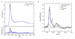
Investigating DNA Radiation Damage Using X-Ray Absorption Spectroscopy
The key to achieving more effective radioprotection and radiotherapy is to understand the exact mechanism of the interaction between radiation and biomolecules, and in particular to obtain the precise structure of the different forms of damage and their relative ratios. Among all biomolecules exposed to radiation, DNA plays an important role because any damage to its molecular structure can affect the whole cell and may lead to chromosomal rearrangements resulting in genomic instability or cell death.
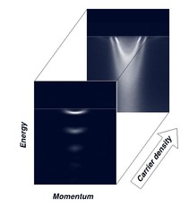
Tailoring Novel Superconductivity
The band insulator strontium titanate SrTiO3 (STO), widely used as a substrate for growing oxide films, is a highly fascinating material. Recently, novel physical properties have been observed at the interface between STO and the materials grown on it. For instance the appearance of superconductivity above the temperature of liquid nitrogen, observed in a single monolayer of FeSe (its critical temperature is higher than in any iron-based bulk material) grown on the STO surface, suggests a key-role of the STO substrate.