
News & Scientific Highlights
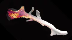
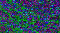
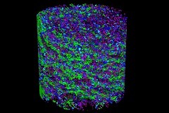
3D nanostructure of a bone made visible
Bones are made up of tiny fibres that are roughly a thousand times finer than a human hair. Researchers at the Paul Scherrer Institute PSI have developed a new computer-based algorithm with which they were able to visualize the localised order and alignment of these nanostructures inside an entire piece of bone for the first time.
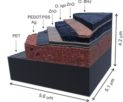
X-ray nanotomography aids the production of eco-friendly solar cells
Polymer solar cells are in the spotlight for sustainable energy production of the future. Characterization of these devices by X-ray nanotomography helps to improve their production using environmentally friendly materials.
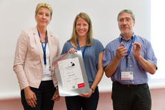
2015 Otto Kratky award
Marianne Liebi was awarded the 2015 Otto Kratky award by the Helmholtz-Centre Berlin for excellence in the field of small-angle X-ray scattering (SAXS) analysis. The award was bestowed in the last SAS2015 conference in Berlin. Marianne is a postdoctoral fellow in the coherent X-ray scattering group (CXS) in PSI, carrying out research in scanning SAXS measurement and analysis in 2D and 3D. Image credit ©HZB/Michael Setzpfandt
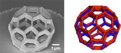
Element-Specific X-Ray Phase Tomography of 3D Structures at the Nanoscale
Recent advances in fabrication techniques to create mesoscopic 3D structures have led to significant developments in a variety of fields including biology, photonics, and magnetism. Further progress in these areas benefits from their full quantitative and structural characterization.
From inside an eggshell
Tiny cavities inside eggshells supply the materials that stimulate and control the shell’s growth. Using a novel imaging technique, researchers from the Paul Scherrer Institute (PSI), ETH Zurich and the Dutch FOM Institute AMOLF have succeeded in depicting these voids in 3D for the first time. In doing so, they lift an old limitation of tomographic images and hope that one day medicine will also benefit from their method.
Multiresolution X-ray tomography, getting a clear view of the interior
Researchers at PSI have developed a technique that combines tomography measurements at different resolution levels to allow quantitative interpretation for nanoscale tomography on an interior region of interest of the sample. In collaboration with researchers of the institute AMOLF in the Netherlands and ETH Zurich in Switzerland they showcase their technique by studying the porous structure within a section of an avian eggshell. The detailed measurements of the interior of the sample allowed the researchers to quantify the ordering and distribution of an intricate network of pores within the shell.
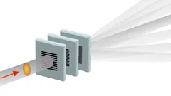
Split x-ray flash shows rapid processes
SwissFEL, PSI’s x-ray laser, is to render the individual steps of very rapid processes visible. A new method will facilitate especially precise experiments: the individual x-ray flashes are split into several parts that arrive at the object under examination one by one. The principle of the method harks back to the ideas of the earliest high-speed photography.
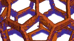
Nanometres in 3D
Scientists at the Paul Scherrer Institute and ETH Zurich have created 3D images of tiny objects showing details down to 25 nanometres. In addition to the shape, the scientists determined how particular chemical elements were distributed in their sample and whether these elements were in a chemical compound or in their pure state.
Innovation Award on Synchrotron Radiation 2014 for high-resolution 3D hard X-ray microscopy
The 2014 Innovation Award on Synchrotron Radiation was bestowed to researchers Ana Diaz, Manuel Guizar-Sicairos, Mirko Holler, and Jörg Raabe from the Paul Scherrer Institut, Switzerland, for their contributions to method and instrumentation development, which have set new standards in high-resolution 3D hard X-ray microscopy.