
Lab News & Scientific Highlights
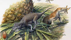
Côté menu, les premiers mammifères avaient déjà leurs préférences
De toutes nouvelles analyses de petits mammifères fossiles livrent de nouveaux éléments sur la connaissance du menu de nos plus lointains ancêtres. Les résultats permettent de comprendre comment sont apparues certaines caractéristiques typiques des mammifères actuels. Ces conclusions reposent sur des analyses, qui ont été réalisées à la Source de Lumière Suisse (SLS) de l’Institut Paul Scherrer, au moyen de la tomographie par rayons X.
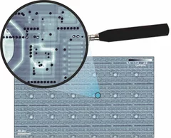
Fast scanning coherent X-ray imaging using Eiger
The smaller pixel size, high frame rate, and high dynamic range of next-generation photon counting pixel detectors expedites measurements based on coherent diffractive imaging (CDI). The latter comprises methods that exploit the coherence of X-ray synchrotron sources to replace imaging optics by reconstruction algorithms. Researchers from the Paul Scherrer Institut have recently demonstrated fast CDI image acquisition above 25,000 resolution elements per second using an in-house developed Eiger detector. This rate is state of the art for diffractive imaging and even on a par with the fastest scanning X-ray transmission instruments. High image throughput is of crucial importance for both materials and biological sciences for studies with representative population sampling.
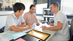
Le contraste de phase améliore la mammographie
Grâce à l'imagerie par contraste de phase, des chercheurs de l’EPF Zurich, de l’Institut Paul Scherrer (PSI) et de l’hôpital cantonal de Baden ont réussi à obtenir des mammographies, grâce auxquelles il est possible de procéder à une évaluation plus précise des cancers et des précancers du sein. Ce procédé pourrait contribuer à une utilisation plus ciblée des biopsies, et à l’amélioration des examens de suivi.
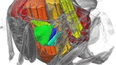
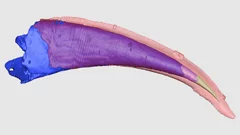
Un film en 3D montre ce qui se passe à l’intérieur d’insectes en plein vol
Grâce aux rayons X produits par la Source de Lumière Suisse SLS, des prises de vue inédites des muscles que les mouches utilisent pour voler (muscles alaires) ont pu être réalisées, à haute vitesse et en 3D. Une équipe de scientifiques de l’Université d’Oxford, de l’Imperial College de Londres et de l’Institut Paul Scherrer (PSI) a développé un procédé de prise de vue tomographique révolutionnaire. Grâce à lui, ils ont pu filmer ce qui se passe à l’intérieur d’insectes en plein vol. Ces films permettent de découvrir en profondeur l’un des mécanismes naturels les plus complexes, et montrent que les déformations structurelles sont la clé pour comprendre la manière dont la mouche contrôle son battement d’aile.

X-ray tomography reaches 16 nm isotropic 3D resolution
Researchers at PSI reported a demonstration of X-ray tomography with an unmatched isotropic 3D resolution of 16 nm in Scientific Reports. The measurement was performed at the cSAXS beamline at the Swiss Light Source using a prototype instrument of the OMNY (tOMography Nano crYo) project. Whereas this prototype measures at room temperature and atmospheric pressure, the OMNY system, to be commissioned later this year, will provide a cryogenic sample environment in ultra-high vacuum without compromising imaging capabilities. The researchers believe that such a combination of advanced imaging with state-of-the-art instrumentation is a promising path to fill the resolution gap between electron microscopy and X-ray imaging, also in case of radiation-sensitive materials such as polymer structures and biological systems.

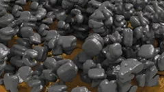
Unique insight into carbon fibers on the nanoscale
The investigation of the mechanical properties of carbon fibers benefits from highly resolved three-dimensional density maps within representative volumes, but such images are not easily obtained with standard methods. Scientists from the Paul Scherrer Institut in collaboration with Honda R&D in Germany have recently visualized density distributions on the sub-micrometer scale within entire carbon fiber sections, revealing surprising graphite distributions within the fibers. This capability will prove useful for the systematic characterization of fibers, contributing to the development of light and robust materials at lower costs.
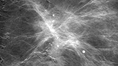
Neues Diagnoseverfahren bei Brustkrebs vielversprechend
Ein neues Mammografie-Verfahren könnte laut einer soeben veröffentlichten Studie einen deutlichen Mehrwert für die Diagnose von Brustkrebs in der medizinischen Praxis bringen. Die am PSI in Zusammenarbeit mit dem Brustzentrum des Kantonsspitals Baden und dem Industriepartner Philips entwickelte Methode macht bereits kleinste Gewebeveränderungen besser sichtbar. Dies könnte potenziell die Früherkennung von Brustkrebs verbessern. Zukünftig sollen Studien an Frauen mit einer Brustkrebserkrankung den Mehrwert der Methode endgültig belegen.Cette actualité n'existe qu'en anglais et allemand.
Mit Röntgenstrahlen der Lebensdauer von Lithium-Ionen-Akkus auf der Spur
Mithilfe von Röntgen-Tomographie haben Forschende die Vorgänge in Materialien von Batterie-Elektroden detailliert untersuchen können. Anhand hochaufgelöster 3D-Filme zeigen sie auf, weshalb die Lebensdauer der Energiespeicher begrenzt ist.Cette actualité n'existe qu'en anglais et allemand.
Zähnen
Mit Hilfe von Röntgenlicht aus der Synchrotron Lichtquelle Schweiz des PSI ist es Paläontologen der Universität Bristol gelungen, ein Rätsel um den Ursprung der ersten Wirbeltiere mit harten Körperteilen zu lösen. Sie haben gezeigt, dass die Zähne altertümlicher Fische (der sogenannten Conodonten) unabhängig von den Zähnen und Kiefern heutiger Wirbeltiere entstanden sind. Die Zähne dieser Wirbeltiere haben sich vielmehr aus einem Panzer entwickelt, der dem Schutz vor den Conodonten, den ersten Raubtieren, diente.