Coherent X-ray Scattering Group (CXS)

The Coherent X-ray Scattering (CXS) group develops techniques in scanning- and time-resolved SAXS and high-resolution scanning X-ray microscopy at the cSAXS beamline. In collaboration with research groups, within PSI and international universities and research institutes, we apply these techniques to a wide range of problems in the fields of biology, biomedical research and materials science.
We will open position at cSAXS for small-angle scattering tensor tomography in combination with ptychographic tomography. Contact us for details.
Scientific Highlights
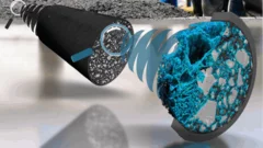
A deep look into hydration of cement
Researchers led by the University of Málaga show the Portland cement early age hydration with microscopic detail and high contrast between the components. This knowledge may contribute to more environmentally friendly manufacturing processes.
Manuel Guizar-Sicairos appointed as Associate Professor at EPF Lausanne and head of the Computational X-ray Imaging group at PSI
Dr. Manuel Guizar-Sicairos, currently Senior Scientist at PSI, was appointed as Associate Professor of Physics in EPF Lausanne and head of the Computational X-ray Imaging group in PSI.
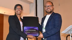
Dr. Manuel Guizar-Sicairos elected as SPIE Fellow member
Dr. Manuel Guizar-Sicairos was elected as a 2022 SPIE Fellow Member for his contributions to coherent lensless imaging, including ptychography and X-ray nano-tomography. The distinction was awarded in the SPIE’s Optics & Photonics conference in San Diego, California.


