
News

High-numerical-aperture optics is key to ultra-fast tomographic microscopy
A novel high-numerical-aperture macroscope optics dedicated to high-temporal and high-spatial resolution X-ray tomographic microscopy is available at TOMCAT. Coupled with the in-house developed GigaFRoST camera, this highly efficient imaging setup enables tomographic microscopy studies at 20 Hz and beyond, opening up new possibilities in tomographic investigations of dynamic processes. A detailed characterization of the macroscope performance was published in Journal of Synchrotron Radiation on May 21, 2019.

From whole organ imaging down to single cell analysis
Researchers from the TOMCAT beamline, University College London (UCL), IDIBAPS and Universitat Pompeu Fabra (UPF) have developed a methodology that allows the multiscale analysis of the structural changes resulting from remodelling cardiovascular diseases, from whole organ down to single-cell level. This methodology has been published as an article in the journal Scientific Reports on May 6th 2019.
Dr. Margie Olbinado joins as Industrial Liaison Scientist
Dr. Margie Olbinado joins the X-ray Tomography group as scientist to take care of industrial tomographic imaging and business strategies for the growing TOMCAT industrial portfolio. Before joining PSI, Margie was a scientist at The European Synchrotron - ESRF in France. As industrial liaison, she will work in collaboration with the PSI Technology Transfer, ANAXAM and SLS TT AG.
PSI Thesis Medal goes to Dr. Matias Kagias
The PSI Thesis Medal is awarded every second year to the best PhD thesis performed at the Paul Scherrer Institut. Matias received the prize for his excellent thesis entitled "Direct Self-Imaging Methods for X-ray Differential Phase and Scattering Imaging". Congratulations!
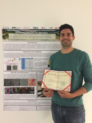
Hector Dejea receives an Outstanding Poster Presentation Award at the bMASR Conference
Hector Dejea, a PhD at TOMCAT, received an Outstanding Poster Presentation Award at the 9th bioMedical Applications of Synchrotron Radiation (bMASR2018) conference held in Beijing (China) from October 23rd till 27th 2018. He presented the latest results of his work, entitled Multiscale X-ray Phase Contrast Imaging for Assessing Cardiac Remodelling: towards in-vitro applications.

TOMCAT paper on hard X-ray multi-projection imaging published
The TOMCAT team in collaboration with scientists from CFEL, MaxIV and ESRF developed a method for hard X-ray multi-projection imaging, using a single crystal to split the beam into multiple beams with different directions.
Dr. Anne Bonnin gives an invited talk at SRI 2018
The 13th International Conference on Synchrotron Radiation Instrumentation (SRI 2018) was hosted by the National Synchrotron Radiation Research Center (NSRRC) from June 10 to 15, 2018. The 5-day conference was gathering scientists and engineers around the world involved in development of new concepts, techniques, and instruments related to synchrotron radiation and free electron laser research.
Arttu Miettinen joins the TOMCAT team as Post Doc
After his PhD at the University of Jyväskylä in Finnland in image analysis, Arttu will be working on the stitching and segmentation of large datasets in the framework of the Human Brain Project.
Dr. Konstantins Jefimovs wins the 2nd poster prize
Dr. Konstantins Jefimovs from the TOMCAT team at SLS was awarded with the 2nd poster prize at the 4th XNPIG Conference (X-ray and Neutron Phase Contrast Imaging with Gratings) held in Zürich, September 12th-15th. He presented latest achievements on gratings fabrication under the title: “Large area small pitch gratings for X-ray interferometry by Displacement Talbot Lithography”.