
Scientific Highlights

High-resolution ptychographic imaging at a seeded free-electron laser source using OAM beams
Electromagnetic waves possessing orbital angular momentum (OAM) are powerful tools for applications in optical communications, quantum technologies, and optical tweezers. Now, a consortium of collaborators in France, Italy, Slovenia, Spain, Switzerland, Sweden, and the US reports on using such beams in the extreme ultraviolet region for ptychographic imaging in the cover page article of Optica 11, Issue 3. By controlling the topological charge, the researchers achieve an improvement of 30% in image resolution.
")
What will the SLS 2.0 upgrade mean for experiments?
Tighter beams, brighter light and extended photon energies open new experimental possibilities.
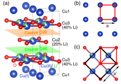
Electron Glass Phase with Resilient Zhang-Rice Singlets in LiCu3O3
LiCu3O3 is an antiferromagnetic mixed valence cuprate where trilayers of edge-sharing Cu(II)O (3d9) are sandwiched in between planes of Cu(I) (3d10) ions, with Li stochastically substituting Cu(II). Angle-resolved photoemission spectroscopy (ARPES) and density functional theory reveal two insulating electronic subsystems that are segregated in spite of sharing common oxygen atoms: a Cu dz2/O pz derived valence band (VB) dispersing on the Cu(I) plane, and a Cu 3dx2−y2/O 2px,y derived Zhang-Rice singlet (ZRS) band dispersing on the Cu(II)O planes.
Developing detectors to transform science with light (part 2)
Part II: Why detecting soft X-rays is hard, and how a new breakthrough is set to transform low energy X-ray science.
Flat-band hybridization between f and d states near the Fermi energy of SmCoIn5
We present high-quality angle-resolved photoemission (ARPES) and density functional theory calculations (DFT+U) of SmCoIn5. We find broad agreement with previously published studies of LaCoIn5 and CeCoIn5, confirming that the Sm 4f electrons are mostly localized. Nevertheless, our model is consistent with an additional delocalized Sm component, stemming from hybridization between the 4f electrons and the metallic bands at “hot spot” positions in the Brillouin zone.
 oxalate solution. The large bandwidth emission covers the full XANES range, making time-consuming monochromator scans obsolete.")
Efficient transient X-ray absorption spectroscopy
By combining the unique large bandwidth emission mode of SwissFEL’s ARAMIS undulator and diffractive X-ray optics made of diamond, we have demonstrated a new method for time-resolved X-ray absorption near edge structure (XANES) spectroscopy that enables faster data acquisition and requires smaller sample quantities for high-quality data.

2024 SPIE Advanced Lithography + Patterning, San Jose, California
2024 SPIE Advanced Lithography + Patterning symposium hosted leading researchers who are solving challenges in optical and EUV lithography, patterning technologies, metrology, and process integration for semiconductor manufacturing and adjacent applications. The symposium features six conference topics.

The Tipping Point!
Exciting to see that some of our research on Narwhal tusk made it into an educational videogame about climate change in the Arctic and its impact on some of its inhabitants!

Charge fractionalisation observed spectroscopically
Quantum mechanics tells us that the fundamental unit of charge is unbreakable – but exceptions exist.

Introduction to Muon Spin Spectroscopy
Alex Amato and Elvezio Morenzoni (both NUM) have published a new textbook entitled 'Introduction to Muon Spin Spectroscopy: Applications to Solid State and Material Sciences'. The book is ideal for a first course in muon spin spectroscopy (µSR), comes enriched with exercises and solutions to master the subject and includes practical examples to quantify key experimental parameters.