
Archive
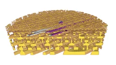
3-D X-ray imaging makes the finest details of a computer chip visible
Researchers at the PSI have made detailed 3-D X-ray images of a commercially available computer chip. In their experiment, they examined a small piece that they had cut out of the chip beforehand. This sample remained undamaged throughout the measurement. It is a major challenge for manufacturers to determine if, in the end, the structure of their chips conforms to the specifications. Thus these results represent one important application of an X-ray tomography method that the PSI researchers have been developing for several years.
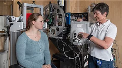
Rays of hope for patients
For over 30 years, patients with a particular form of ocular tumour have been treated at PSI by means of proton irradiation. The tiny particles hit their target with millimetre precision, without endangering other structures of the eye. The irradiation facility OPTIS, developed at the PSI Center for Proton Therapy of the PSI, is a success story, considering that for more than 90 percent of the patients treated to date, the eye could be saved.
