
Lab News & Scientific Highlights
Dr. Manuel Guizar-Sicairos elected as Fellow member of The Optical Society (OSA)
Dr. Manuel Guizar-Sicairos, beamline scientist at the cSAXS beamline, was elected as a Fellow Member of The Optical Society (OSA) for seminal contributions to methods and applications of coherent lensless imaging, ptychography, x-ray nanotomography, and new modalities of x-ray microscopy.
Measurements at PSI enabled detailed understanding of genetic scissors
PSI congratulates Emmanuelle Charpentier and Jennifer Doudna on winning this year's Nobel Prize in Chemistry. Experiments at the Swiss Light Source SLS in 2013 made it possible to elucidate the structure of the protein complex CRISPR-Cas9.
A question of binding
At PSI, researchers are screening molecule fragments to see if these bind to important proteins of the coronavirus SARS-CoV-2 and thus have the potential to disable it. They are hoping the many individual pieces of information will yield an answer as to what an effective drug might look like.

3D printing silica aerogels at the micrometer scale
A group of EMPA and ETH Zürich researchers have developed a new method to directly write ink made of silica aerogels in 3D. Thanks to X-ray phase contrast tomography at the TOMCAT beamline they characterized the resulting printed material with different compositions. Their results were published in Nature on August 18, 2020.
")
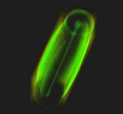
Phase contrast microtomography reveals nanoparticle accumulation in zebrafish
Metal-based nanoparticles are a promising tool in medicine – as a contrast agent, transporter of active substances, or to thermally kill tumor cells. Up to now, it has been hardly possible to study their distribution inside an organism. Researchers at the University of Basel in collaboration with the TOMCAT team have used phase contrast X-ray tomographic microscopy to take high-resolution captures of the nanoparticle aggregation inside zebrafish embryos.
The study was published in the journal Small and featured on the cover of its current issue.
Covid-19 research: Anti-viral strategy with double effect
Frankfurt scientists identify a possible weakness of the SARS-CoV-2 virus. They carried out part of their measurements at PSI's Swiss Light Source SLS. The research results are published this week in the scientific journal Nature.

First MX results of the priority COVID-19 call
The Dikic group at the Goethe University in Frankfurt am Main, Germany has published the first results following the opening of the "PRIORITY COVID-19 Call” at SLS.
Miniaturized fluidic circuitry observed in 3D
The team of Prof. Thomas Hermans at the University of Strasbourg in France managed to create wall-less aqueous liquid channels called anti-tubes. Thanks to X-ray phase contrast tomography at the TOMCAT beamline those anti-tubes could be observed in 3D. The exciting results were published in Nature on May 6, 2020.

Microcalcifications in breast tissue: Paving the way for future diagnostic solutions?
Microcalcifications are a common sign in mammography. For example, in 90% of ductal carcinoma in situ microcalcifications are the first and unique sign indicating the presence of the lesion. Nevertheless, up to around 50% of the resulting biopsies reveal a benign lesion, a 'false positive'. Researchers were able to show now that breast microcalcifications detected in tumors have specific chemical and crystalline features different from those observed in benign samples. Moreover, microcalcifications detected in tumors but located outside the lesion show malignant features too. This indicates that cancer influences the surrounding tissue even if it exhibits apparently healthy morphological features. These results indicate that the biochemical differences between benign and malignant microcalcifications can be potentially identified by light-based tools, able to investigate microcalcifications inside the breast without performing biopsies.