  • Paul Scherrer Institut PSI
  • PSI Research, Labs & User Services

Digital User Office

  • Digital User Office
  • DE
  • EN
  • FR
Paul Scherrer Institut (PSI)
Suche
Paul Scherrer Institut (PSI)

Hauptnavigation

  • Research at PSIOpen mainmenu item
    • Research Initiatives
    • Research Integrity
    • Scientific Highlights
    • Scientific Events
    • Scientific Career
    • PSI-FELLOW
    • PSI Data Policy
  • Research Divisions and LabsOpen mainmenu item
    • Overview
    • Research with Neutrons and Muons
    • Photon Science
    • Energy and Environment
    • Nuclear Energy and Safety
    • Biology and Chemistry
    • Large Research Facilities
  • Facilities and InstrumentsOpen mainmenu item
    • Overview
    • Large Research Facilities
    • Facilities
    • PSI Facility Newsletter
  • PSI User ServicesOpen mainmenu item
    • User Office
    • Methods at the PSI User Facilities
    • Proposals for beam time
    • Proposal Deadlines
    • Data Analysis Service (PSD)
    • EU support programmes
  • DE
  • EN
  • FR

Digital User Office (mobile)

  • Digital User Office

You are here:

  1. PSI Home
  2. Labs & User Services
  3. PSD
  4. LMN
  5. Research Groups
  6. SwissFEL nanoengineering
  7. Bio-Interfaces

Secondary navigation

Laboratory for Micro and Nanotechnology

  • About LMN
    • Organisational Structure
  • Open Positions
  • People
  • Research Groups Expanded submenu item
    • Nanotechnology
    • X-ray Optics and Applications
      • X-ray Optics for Imaging and Spectroscopy
        • Fresnel Zone Plate for X-ray Microscopy
        • Blazed X-ray Optics
        • Zernike X-ray Phase Contrast Microscopy
        • Fresnel Zone Plates for RIXS
        • Refractive Lenses by 2 Photon 3D Lithography
      • Wavefront Metrology and Manipulation
        • Vortex Fresnel Zone Plates
        • Grating-based Wavefront Metrology
      • X-ray Optics for XFELs
        • Diamond Fresnel Zone Plates
        • Beam Splitter Gratings for Spectral Monitoring
        • A Delay Line for Ultrafast Pump-Probe Experiments
        • X-ray Streaking for Ultrafast Processes
    • Polymer Nanotechnology
      • Nanoimprint Lithography
      • Three Dimensional Structures
    • Molecular Nanoscience
      • On-surface Chemistry
      • Spins in Molecular Monolayers
      • SiC: Surfaces and Interfaces
      • Our Research Team
    • Advanced Lithography and Metrology
      • EUV Interference Lithography
      • EUV Lensless Imaging
      • ALM Nanoscience
    • Quantum Technologies
      • News and highlights
      • People
      • Open positions
      • Current projects
        • 2D semiconductor devices
        • CDW-based memory devices
        • Imaging quantum many-body states
        • Nonlinear magnonics
        • Rare-earth quantum magnets
        • Strained Germanium laser
      • Techniques
        • Cristallina-Q
        • IR beamline
        • Nano-fabrication
      • Publications
      • QTC@PSI
  • Facilities and Equipment
    • Cleanroom Labs
    • Surface Science Lab
    • Scanning Electron Microscopy
    • Scanning Probe Microscopy
    • PEARL Beamline
    • XIL Facility at the SLS
    • Nanoimprint Facilities
    • Electron Beam Lithography
  • LMN News
  • LMN Highlights
    • Archive
  • Publications
    • Publications 2011 - 2016

Biointerfaces: Enabling Technology for Bio-Research

In collaboration with research groups from PSI and Universities we are developing surfaces for application in biomolecular and biomaterials research ranging from protein patterns on wafer, glass and polymer surfaces to micro- and nanostructured surfaces and templates for replication in biocompatible polymers.


Specific projects

Neuron cells cultured on a structured substrate
Outgrowth of neurites from primary rat neurons guided by a structure of parallel ridges.

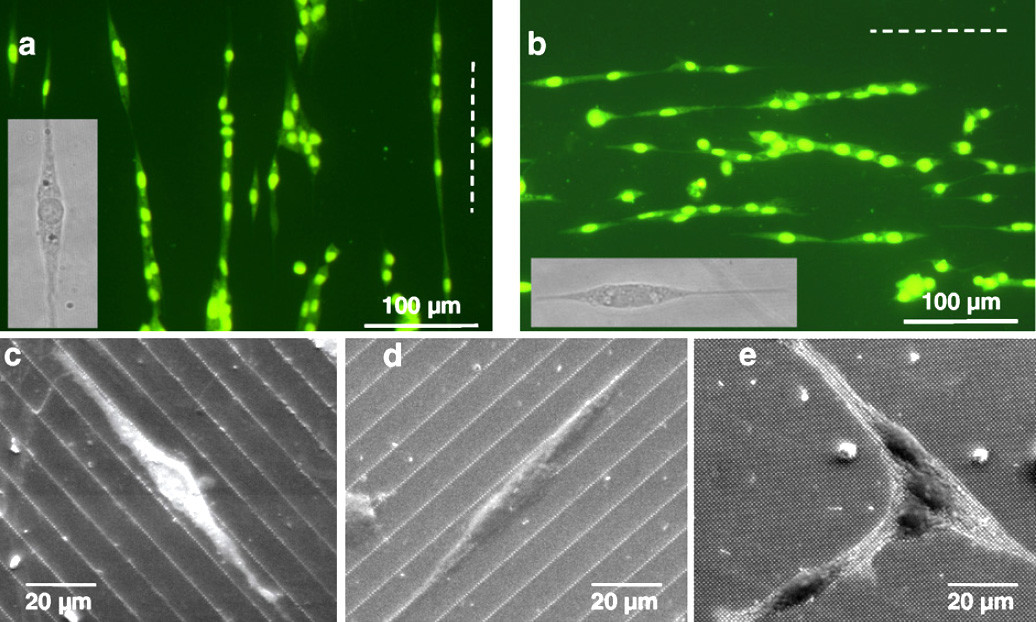
Substrates for ultrastructural studies     We are developing patterned supports for neuron cell co-cultures with the aim to replicate the neuronal pathway under controlled conditions for subsequent use in ultrastructural studies of Parkinson’s Disease (PD). The lithographically structured substrates are compatible with a cryo-ultrastructural analysis workflow consisting of high pressure freezing, freeze substitution and preparation for electron microscopy. The aim is to understand the ultrastructure of PD-associated sub-cellular aggregates and intracellular components within diseased synapses, axons and neuronal bodies, in order to design better therapeutic strategies and ways to potentially monitor and detect the disease at an early stage. Our well-designed cell culture model can provide key insights while circumventing complex issues associated with handling whole human brain tissue.


Functionalized microneedle integrated into an optofluidic sensor device

Integrated microneedle-optofluidic biosensor     In an international collaboration between the University of the British Columbia (UBC) and the Paul Scherrer Institut (PSI), a promising system for painless and minimally-invasive therapeutic drug monitoring has been demonstrated. The proposed device is based on the combination of an optofluidic system with hollow microneedles to extract extremely small volumes (< 1 nL) of interstitial fluid (ISF) to measure drug concentrations.  >>read more


Alignment of PAE cells cultured on a pattern of VEGF

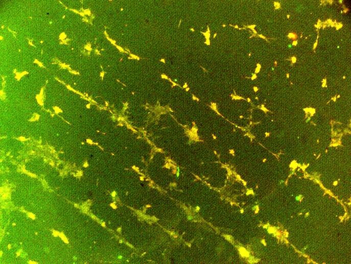
VEGF-Patterns     Based on photolithograpghy or on microfluidics we were producing patterns of proteins of the VEGF-Family. Studies of porcine aortic endothelial cells (PAE cells) cultured on the substrates enable new insights into the mechanisms deteriming blood vessel formation which are highly dependent on the presence and distribution of VEGF-proteins. In collaboration with Kurt Ballmer, molecular cell biology at PSI.


Growth of NSCs on pillar arrays replicated in biocompatible PLLA/PLGA-copolymers. The cells align along the most dense packing of nanopillars.
Nanopillar arrays for cell growth studies    In a collaboration with the "Biomaterials and Tissue Engineering Research Laboratory" (BIOMAT), METU, Ankara, we designed and produced of arrays of 200 nm to 1 µm wide and 1 to  5 µm tall pillars with pillar-to-pillar distances in the range of 1-10 µm. The pillar arrays were replicated into biocompatible polymers and are being used in cell growth experiments performed at METU. Studies of cell adhesion and proliferation of different cells depending on the pillar size and inter-pillar distances deliver valuable information for the design of medical implant surfaces.


Sidebar

Contact

Dr. Celestino Padeste

Division of Biology and Chemistry
Paul Scherrer Institut
Forschungsstrasse 111
5232 Villigen PSI
Switzerland

Telephone:
+41 56 310 2141

E-mail:
celestino.padeste@psi.ch

top

Footer

Paul Scherrer Institut

Forschungsstrasse 111
5232 Villigen PSI
Switzerland

Telephone: +41 56 310 21 11
Telefax: +41 56 310 21 99

How to find us
Contact form

Visitor Centre psi forum
School Lab iLab (in German)
Center for Proton Therapy
PSI Education Centre
PSI Guest House
PSI Gastronomie (in German)


Service & Support

  • Phone Book/People Search
  • User Office
  • Accelerator Status
  • PSI Publications
  • Suppliers
  • E-Billing
  • Computing
  • Safety (in German)

Career

  • Working at PSI
  • Job Opportunities
  • Training and further education
  • Vocational Training (in German)
  • PSI Education Center

For the media

  • PSI in brief
  • Facts and Figures
  • Media Contact
  • Media Releases
  • Social Media Newsroom

Follow us: Twitter (in English) LinkedIn Youtube Issuu RSS

Footer legal

  • Imprint
  • Terms and Conditions
  • Editors' login