
Lab News & Scientific Highlights

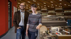
SLS MX beamtime update
Update of the SLS MX beamline operation during the COVID-19 period

X-ray Imaging for Biomedicine: Imaging Large Volumes of Fresh Tissue at High Resolution
The TOMCAT beamline at the Swiss Light Source specializes in rapid high-resolution 3-dimensional tomographic microscopy measurements with a strong focus on biomedical imaging. The team has recently developed a technique to acquire micrometer-scale resolution datasets on the entire lung structure of a juvenile rat in its fresh natural state within the animal’s body and without the need for any fixation, staining or other alteration that would affect the observed structure (E. Borisova et al., 2020, Histochem Cell Biol).

Nanowelten in 3-D
Tomogramme aus dem Inneren von Fossilien, Hirnzellen oder Computerchips liefern neue Erkenntnisse über feinste Strukturen. Die 3-D-Bilder gelingen mithilfe der Röntgenstrahlen der Synchrotron Lichtquelle Schweiz SLS dank eigens entwickelter Detektoren und raffinierter Computeralgorithmen.
Kurzfilm eines magnetischen Nanowirbels
Mit einer neu entwickelten Untersuchungsmethode konnten Forschende die magnetische Struktur im Inneren eines Materials mit Nanometer-Auflösung abbilden. Ihnen gelang ein kurzer «Film» aus sieben Bildern, der erstmalig in 3-D zeigt, wie sich winzige Wirbel der Magnetisierung tief im Inneren eines Materials verändern.

Innovation Award on Synchrotron Radiation 2019 for the development of XFEL detectors using the adaptive gain principle
The Innovation Award on Synchrotron Radiation 2019 was given to the researchers Prof. Heinz Graafsma from Desy and Dr. Aldo Mozzanica and Dr. Bernd Schmitt both from the Paul Scherrer Institute. The three physicists were honored for their contributions to the development of detectors for XFEL applications based on the dynamic gain switching principle enabling simultaneously single photon resolution and a large dynamic range. The laudation was held by Prof. Edgar Weckert from Desy. The Synchrotron Radiation Innovation Award is sponsored by SPECS GmbH and BESTEC GmbH.

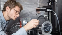
MacEtch in gas phase: a new nanofabrication technology at PSI
The grating fabrication team of the X-ray tomography group has scored another record in etching technology of silicon by realizing a MacEtch process in gas phase. Ultra-high aspect ratios (up to 10 000 : 1) in the nanoscale regime (down to 10 nm) were achieved by platinum assisted chemical etching of silicon in the gas phase. The results were published in Nanoscale Horizons on February 17, 2020.
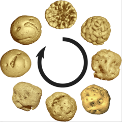
Animal embryos evolved before animals
Detailed characterization of cellular structure and development of exceptionally preserved ancient tiny fossils from South China by synchrotron based X-ray tomographic microscopy at TOMCAT led an international team of researchers from the University of Bristol and Nanjing Institute of Geology and Palaeontology to the discovery that animal-like embryos evolved long before the first animals appear in the fossil record.
Faserverstärkte Verbundstoffe schnell und präzise durchleuchten
Forschende des Paul Scherrer Instituts PSI haben eine neues Verfahren entwickelt, mit dem sich faserverstärkte Verbundwerkstoffe präzise durchleuchten lassen. Das könnte helfen, bessere Materialien mit neuartigen Eigenschaften zu entwickeln.
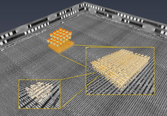
3D imaging for planar samples with zooming
Researchers of the Paul Scherrer Institut have previously generated 3-D images of a commercially available computer chip. This was achieved using a high-resolution tomography method. Now they extended their imaging approach to a so-called laminography geometry to remove the requirement of preparing isolated samples, also enabling imaging at various magnification. For ptychographic X-ray laminography (PyXL) a new instrument was developed and built, and new data reconstruction algorithms were implemented to align the projections and reconstruct a 3D dataset. The new capabilities were demonstrated by imaging a 16 nm FinFET integrated circuit at 18.9 nm 3D resolution at the Swiss Light Source. The results are reported in the latest edition of the journal Nature Electronics. The imaging technique is not limited to integrated circuits, but can be used for high-resolution 3D imaging of flat extended samples. Thus the researchers start now to exploit other areas of science ranging from biology to magnetism.